
Have a question about one of our products? Check out our technical library for recently asked questions from other scientists around the world.
Technical Library
Recent Entries in Technical Library
299 Entries on 3 Pages
-
We use Macrophage Generation Media from PromoCell to get differentiated macrophages from peripheral blood monocytes. The monocytes are isolated by culturing PBMCs in Monocyte Attachment Media (also from PromoCell) for 1.5 h. Would it be ok if the attachment step is done overnight, since we receive the blood later in the day which makes it difficult to process on the same day?
It is not recommended to leave the blood cells in the Monocyte Attachment Medium for longer than 1.5-2 hrs. The medium was developed for (short-term) attachment of the monocytes and does not provide nutrients for a longer time period. Leaving the cells in Monocyte Attachment Medium for a longer time or even overnight will induce apoptosis and lead to the loss of the cells.
If necessary, you can reduce the incubation time to 1 hr. In this case, it is advisable to equilibrate the media in the incubator before so that you can immediately and directly add the appropriate amount of PBMC suspension.Related Links and Documents
-
Does PromoCell recommend a particular manufacturer or brand of tissue culture plastic to grow the primary cells or can I use any supplier on the market?
Our customers have successfully used TC flasks and dishes from all the leading cell culture plastic suppliers to grow PromoCell’s primary human cells. We do not have any knowledge whether the dishes from local TC plastic suppliers work in the same way. We recommend to first test whether these brands provide the same good performance as the plastic of the leading manufacturers.
-
After differentiation of human MSC into mature adipocytes – what medium do I have to use to culture those cells?
Differentiation of hMSC into mature adipocytes takes approx. 2 weeks. You can keep the adipocytes for up to 3 weeks in the MSC Adipogenic Differentiation Medium. After 3 weeks we recommend to switch to PromoCell’s Adipocyte Nutrition Medium (C-27438).
Related Links and Documents
-
What is the composition of Melanocyte Growth Medium M2 SupplementMix (C-39420)?
Supplement Mix for Melanocyte Growth Medium M2 (C-39420) is free of serum and doesn’t contain any PMA. The qualitiative and quantitative composition is proprietary.
Related Links and Documents
-
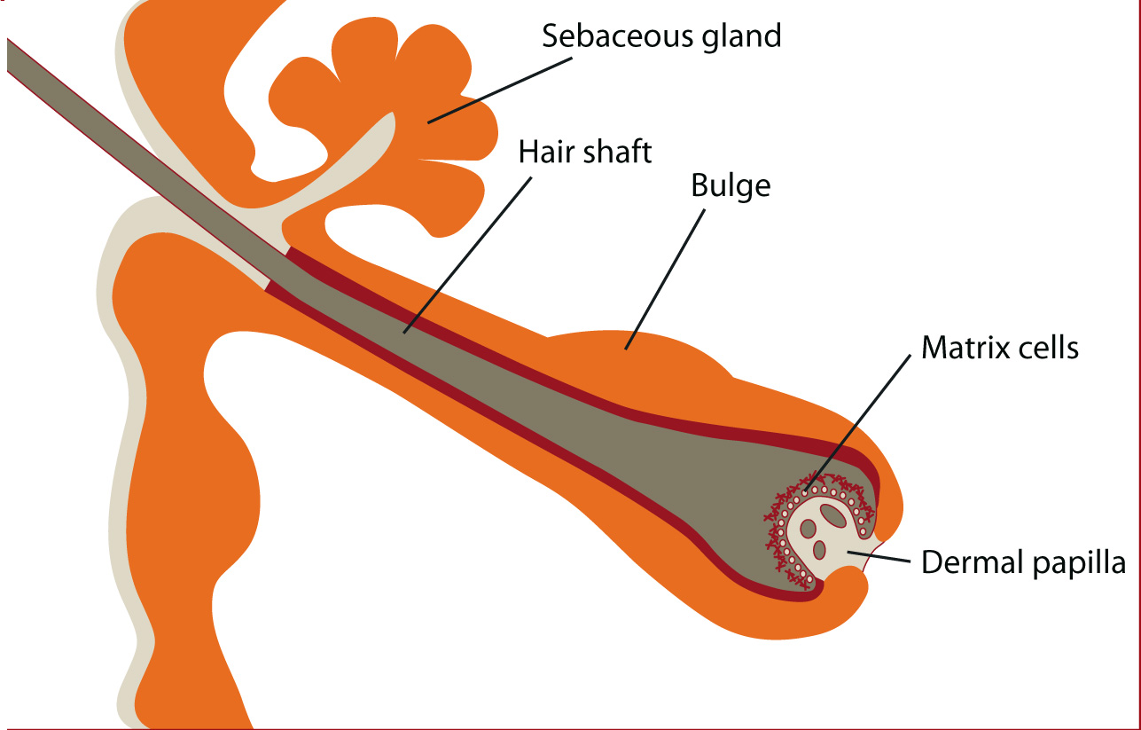
What is the source of PromoCell’s Normal Human Follicle Dermal Papilla Cells? For how long can they be maintained in culture?
HFDPC are isolated from the hair papilla of normal human scalp hair follicles. Hair papilla in the adult hair follicle play a crucial role in the dermal-epidermal interactions that control hair production and in hair growth cycle events. The follicle dermal cells are cryopreserved at second passage and can be cultured for at least 10 population doublings when using PromoCell Follicle Dermal Papilla Cell Growth Medium (Cat. C-26501). Typical population doubling times are between 20-36 hrs.
The recommended seeding density after thawing/trypsinization is 5,000-10,000 cells/cm2. Using 1:4 splits, you can perform 4-5 passages with the cells.Related Links and Documents
-
Do PromoCell HDMEC exhibit migration and tube formation as a response to angiogenic stimuli?
In principle, all microvascular endothelial cells should be able to migrate, proliferate and form tubes or sprouts in an appropriate assay after angiogenic stimulation. As this is not part of our routine quality control procedure, we cannot tell for sure whether all cell lots will respond to angiogenic stimuli. However PromoCell also supplies HDMEC pre-screened (C-12215) that are especially tested for a positive VEGF response.
Related Links and Documents
-
How should I store the PromoCell media and supplements?
Upon arrival, the basal media should be stored between 4°C and 8°C, the supplements at -20°C.
-
How does PromoCell characterize their kidney cells (HREpC; HRCEpC) ?
Our kidney cells are characterized by their epithelial cell morphology and by cytokeratin expression using a pan-cytokeratin antibody.
Related Links and Documents
-
Which medium do PromoCell recommend to use on HUVEC – Endothelial Cell Growth Medium or Endothelial Cell Growth Medium 2?
The standard medium for isolation and propagation of our HUVEC, HUAEC, HPAEC, and HSaVEC is Endothelial Cell Growth Medium (C-22010). It contains ECGS, an extract from bovine hypothalamus which has mitogenic effects on endothelial cell proliferation. Scientists who prefer a more defined Growth Medium can use Endothelial Cell Growth Medium 2 (C-22011). In this medium, ECGS is replaced by VEGF, IGF, and additional bFGF and EGF to stimulate endothelial cell growth.
Related Links and Documents
-
Can I use my own trypsin or other commercially available trypsin solutions for subculturing PromoCell Normal Human Cells?
Most commercially available trypsin solutions have trypsin concentrations of 0.05% or higher which can harm the primary cells. PromoCell recommend using a 0.04% trypsin / 0.03 % EDTA solution for the subculture of Normal Human Cells.
Related Links and Documents
-
What plating density should I use for PromoCell Human Adult Stem and/or Blood Cells?
The recommended plating density after thawing/subculture may vary depending on the cell type. It is specified on the Website (tab: additional Information) and in the Product Manual.
Related Links and Documents
-
How can I test whether my cells are infected with mycoplasma?
Several different methods for the detection of mycoplasmas have been described, like e.g., cultures on agar, in liquid or semi-solid media, staining with DAPI, mycoplasma-specific antibodies, biochemical methods, and PCR-based assays. PCR-based detection is very sensitive, detects all mycoplasma species that occur in cell cultures and is completed within 3-5 hours.
-
Can PromoCell supply preadipocytes (subcutaneous, visceral) from donors with obese BMI / non-smokers / non diabetics? Can PromoCell supply preadipocytes from different age groups?
The basic information we receive from the surgeons about the tissue donors usually includes age, gender, and ethnicity.
- For many of our donors of subcutaneous preadipocytes, we also have information on BMI, hair color, skin pigmentation, and, in some cases, smoking habits or known diseases (e.g. Diabetes). Most of our subcutaneous HWP donors are between ∼20-65 years old.
- The visceral HWP donors are mostly between ∼20-85 years old. For many cell lots we know the BMI, in some cases also the hair color, skin pigmentation, smoking habits, and/or known diseases (e.g. Diabetes or COPD).
If you are looking for particular specifications, please contact our Scientific Support, so that we can offer you appropriate HWP lots.
Related Links and Documents
-
Which type of endothelial cell is best suited for studying angiogenesis?
The study of angiogenesis has been significantly advanced by the ability to culture endothelial cells in vitro. Initially, large vessel ECs, such as those isolated from the human umbilical vein (HUVEC) were used for these studies but increasingly it has been recognized that microvascular endothelial cells are a more appropriate model since angiogenesis involves microvessels rather than large vessel ECs.
-
If I isolate fresh CD14-monocytes for M1/M2 macrophage generation, can I culture them for a period of time before differentiation?
This is not advised. Please seed the freshly isolated CD14-monocytes immediately in the Monocyte Attachment Medium. Adding a culturing step will change the biological characteristics of monocytes very rapidly.
Related Links and Documents
-
Can the medium for M2 macrophages be switched into RPMI or RPMI + M-CSF after differentiation?
Unfortunately, we did not test this in our hands, and it must be tested by the customer. In fact, our medium is completely different from RPMI and therefore we cannot predict if this is working. We only know the successful long-term culture from our system with our media.
Related Links and Documents
-
Are there any differences in the cultivation protocol when cultivating MSCs compared to other cell types?
Yes, there are a few differences:
– We recommend replacing the MSC Growth Medium XF (C-28019) 3-4 h after seeding, as opposed to 16-24 hours after seeding for most other cell types/growth media, including MSC Growth Medium 2.
– When MSC Growth Medium XF, MSC Neurogenic Differentiation Medium, MSC Adipogenic Differentiation Medium 2 or MSC Osteogenic Differentiation Medium are used, flasks have to be coated with 10 µg/cm² (human or bovine) fibronectin according to the instruction manual.
– We strongly recommend using Accutase (C-41310) for cell detachment instead of Trypsin. If Trypsin is used, contact time should not exceed 2 min.Related Links and Documents
-
Can I use a serum-free medium as negative control in the chondrogenic MSC differentiation? I would like to replace the recommended DMEM+10% FCS.
You could probably use our MSC Chondrogenic Differentiation Medium without inducers (C-28014). It is also serum-free and the same as C-28012, just without chondrogenic inducers.
Related Links and Documents
-
If I isolate tumor cells from my PDX animal model using your PCCS, will the animal cells survive?
In our Primary Cancer Culture System (PCCS, C-28081), only the malignant tumor cells can survive. Therefore, benign animal cells (e.g., fibroblasts) will be depleted and the malignant human tumor cells will survive.
Related Links and Documents
-
Is there is a difference in the percentage of differentiated DCs depending on whether we start with cryopreserved or fresh isolated monocytes?
There is no significant difference in the percentage of cells that will differentiate into Dendritic Cells. But when using cryopreserved cells, the initial cell loss will be higher compared to when fresh cells are used. i.e. the final number of differentiated cells that can be expected will be higher with fresh cells as a starting material due to lower cell death rate.
Related Links and Documents
-
Can PromoCell release the species the following cytokines were produced in: basic Fibroblast Growth Factor and Epidermial Growth Factor included in the Endothelial Cell Growth Media supplements?
Both cytokines are produced in E.coli.
Related Links and Documents
-
What is the differentiation ratio that can be achieved with PromoCell’s subcutaneous HWP?
Most of our subcutaneous preadipocyte lots achieve > 80-90% differentiation when differentiation is induced at P2 (directly after thawing).
We generally recommend using cells for differentiation tests that haven’t undergone more than 4-5 doublings (a maximum of 1 passage after thawing), as the differentiation ratio will decline with the age of the cells.Related Links and Documents
-
The HDMEC I purchased from PromoCell seem to contain 2 different cell morphologies. How is this possible? What’s the purity of HDMEC cultures guaranteed by PromoCell?
Our HDMEC are isolated from the dermis of juvenile foreskin or adult skin. The purity is > 95%.
Since the dermis contains blood and lymphatic capillaries, HDMEC cultures comprise blood and lymphatic microvascular endothelial cells that have differing morphologies. Both cell types have a common origin and can be identified by several markers. The ratio of lymphatic and blood derived endothelial cells can vary from lot to lot and is not determined at PromoCell.Related Links and Documents
-
What is the basis formula for PromoCell’s Osteoblast Basal Medium (C-27010)?
PromoCell’s Osteoblast Basal Medium is an optimized media formulation developed for human osteoblast culture. The exact composition is proprietary.
Related Links and Documents
-
Can I expand the human Mesenchymal Stem Cells prior to differentiating them?
These cells are frozen at the end of 2nd culture. Thawing and seeding results in passage 2 (3rd culture). We recommend that they be used for differentiation experiments not later than passage 5.
The differentiation potential of hMSC in vitro is reduced with ongoing population doublings, meaning the earlier differentiation is induced, the higher the differentiation rates.Related Links and Documents
-
What is the lead time when ordering customized media from PromoCell?
The lead time is usually 4-8 weeks.
-
Where in the lung are the HPMEC harvested from? Are the cells from arterioles or capillaries?
Our human pulmonary microvascular endothelial cells (C-12281) are sourced from the lung parenchyma with all large vessels being removed beforehand. Therefore, most of the HPMEC originate from capillaries.
Related Links and Documents
-
Do the PromoCell HCMEC represent the endocardial cells, i.e. those lining the ventricles? If so, why are they referred to as microvascular endothelial cells?
Our HCMEC (Human Cardiac Microvascular Endothelial Cells) are not endocardial cells. They are isolated from the capillaries in the heart muscle. Therefore, they are in fact microvascular.
Related Links and Documents
-
Can I use PBS instead of HepesBSS to wash the cells before trypsinization?
The PromoCell trypsin 0.04% / EDTA 0.03% solution and the PromoCell TNS solution are both based on HepesBSS. Therefore, it is best to also use HepesBSS to wash the cells prior to trypsinization.
Related Links and Documents
-
What plating density should I use for PromoCell Normal Human Cells?
The recommended plating density after thawing/subculture may vary depending on the cell type. Please refer to the data sheet for your cells under “Specifications”.
-
How can I successfully isolate HUVEC from umbilical cords without using antibiotics?
At PromoCell, we get the umbilical cords from our tissue suppliers with no addition of buffers or media. This method prevents the microorganisms from being washed into the blood vessels. Before we start the cell preparation, the umbilical cord is also cut on both ends with a sterile scalpel to provide sterile intersections in addition to the sterile lumen.
This method allows us to isolate sterile endothelial cells from umbilical vein and to plate them in antibiotics-free culture media.Related Links and Documents
-
How does PromoCell recommend subculturing the chondrocytes (e.g. from T25 into T75 flasks, or into petri dishes)? Does PromoCell recommend a specific type or brand?
We recommend a seeding density for chondrocytes between 10,000 and 20,000 cells/cm². This means that a subconfluent T25-flask with approx. 900,000 cells/T25 flask (36,000 cells/cm² ) may be either split into 3 new T25 or seeded in one T75 flask or in one 100 mm petri dish. We do not recommend a specific type or brand for the culture of HCH.
Related Links and Documents
-
What culture conditions are required for culturing PromoCell Normal Human Cells in the respective PromoCell media?
PromoCell Normal Human Cells should be cultured in the appropriate medium at 37°C and 5% CO2 in a humidified atmosphere.
Please note: If using cell culture flasks w/o filter cap, unscrew the cap by half a turn to allow sufficient ventilation. -
Can you please provide me a protocol for the expansion of CD34+ Progenitor Cells in PromoCell Expansion Medium XF?
Short protocol:
- Thaw the cells (C-12921) for 2 min in a 37°C waterbath. Dilute in 9 ml of complete HPC Expansion Medium XF (+ Cytokine Mix E) and count the cells
- Spin down for 10 min at 240xg, aspirate the supernatant, resuspend the pellet at 20,000 cells/ml HPC Expansion Medium XF
- Plate in an appropriate suspension culture vessel and incubate the culture for 2-3 days at 37°C and 5% CO₂
- Then double the media volume by adding fresh complete medium, e.g., 4 ml suspension culture + 4 ml fresh medium (= 8 ml)
- Incubate the cells for an additional 10-12 days by performing a partial medium change every 2-3 days
Example partial medium change: For a culture volume of 8 ml, spin down the cells, aspirate and discard 4 ml of the supernatant, resuspend the cells and add 12 ml of fresh complete medium (= 16 ml).
In combination with the Cytokine Mix E, the HPC Expansion Medium XF typically promotes a 300-1,000 fold expansion of the total cell population. After 2 weeks of expansion about 20-30% of the population express CD34+, indicating a 50-200 fold expansion of CD34+ progenitor cells.
Related Links and Documents
-
Why does the macrophage differentiation in the PromoCell protocol take 10 days? In the literature or following the feedback from other researchers, it usually only takes 7 days.
M1 / M2 polarization also takes seven days in the PromoCell system but the protocol contains two more days for optional macrophage activation. If you only want non-activated M1 / M2 macrophages, the process is usually completed after 7 days.
Nevertheless, PromoCell does not recommend shortening the 10-day protocol because you actually get a plus in viability and cell yield (due to the re-attachment of floating cells) on day 8-10 due to the media change.Related Links and Documents
-
How can I avoid precipitates when preparing my Mesenchymal Stem Cell Adipogenic Differentiation Medium 2?
The supplement should be at room temperature when added to the MSC Adipogenic Basal Medium 2. It may also be beneficial to invert the tube a few times to bring precipitates back into solution.
Please note: It is not recommended to filter the basal medium, supplements, or complete medium, as components that induce or promote differentiation may be removed, resulting in a low differentiation rate when using the medium.
Related Links and Documents
-
We use Macrophage Generation Media from Promocell to get differentiated macrophages from fresh PBMC. Would it be okay if the attachment step with the Monocyte Attachment Medium is done overnight instead of 1.5 h?
We strongly advise against overnight incubation.
The Monocyte Attachment Medium does not contain cytokines/survival factors for the monocytes. If the cells remain in the Monocyte Attachment Medium for longer than 2 hours, they will go into apoptosis and die. The lymphocytes will survive longer. Some of them will attach after such a long time and then cannot be washed off.
Therefore, overnight attachment is absolutely inappropriate. However, if time is short, monocyte attachment can be shortened to 1 hour. To do this, it is best to add Monocyte Attachment Medium to the culture vessels in advance and equilibrate it in the incubator. Remove the vessels from the incubator only briefly to add the appropriate amount of concentrated PBMC suspension. Then, even after 1 hour, most of the monocytes are attached.
Related Links and Documents
-
Can the media from your cancer media toolbox be used for cells of other, non-human species, e.g., from mice?
Yes, our cancer media (Primary Cancer Culture System/PCCS, 3D Tumorsphere Medium XF, Cancer Cell Line Medium XF) also support the growth of murine tumor cells.
Related Links and Documents
-
Do you know in what media the monocyte-derived DCs can be maintained in culture after the differentiation is completed?
You should use complete DC Generation Medium/DC Generation Medium XF (with all the cytokines). As cells are metabolically active, media should be changed every 3 days. We have observed that the dendritic cell phenotype remains stable for up to 7 days.
Related Links and Documents
-
Does Mesenchymal Stem Cell Growth Medium XF (C-28019) contain Phenol Red?
Yes, our MSC Growth Medium XF contains phenol red. The concentration is confidential.
Related Links and Documents
-
What is the source of the trypsin from the Detach Kit (C-41200/C-41210/C-41220)?
The source is porcine pancreas.
Related Links and Documents
-
Why don’t PromoCell specify in the instructions how long the primary cells should be trypsinized?
The time needed to detach our primary cells depends on many different factors like the cell type, cell density, lot #, trypsin concentration, the efficiency of the washing step before adding the trypsin and the trypsinization temperature.
For most cell types we recommend trypsinization at room temperature and direct observation of detachment under the microscope. This way, you can find out your individual trypsinization time and keep the contact time between cells and trypsin to a minimum. Most cells detach after 2-8 min.Please refer to the instructions in the Manual. For some cell types, trypsinization at 37°C or the use of Accutase or another Detachment Solution is recommended.
Related Links and Documents
-
What is the exact source of PromoCell’s subcutaneous and visceral preadipocytes (HWP)?
Our subcutaneous HWP are isolated from subcutaneous fat of different localizations, e.g. abdomen, breast, or upper arm.
The visceral preadipocytes are isolated from fat surrounding e.g. the pericardium, or from the omentum or mediastinum.The exact localization is specified in the Certificate of Analysis. If you need HWP from a particular localization, please contact our Technical Customer Support prior to placing your order.
Related Links and Documents
-
Are the Renal Epithelial Cells isolated from proximal or distal tubuli?
PromoCell provides two types of Renal Epithelial Cells: Human Renal Epithelial Cells (HREpC) and Human Renal Cortical Epithelial Cells (HRCEpC).
HREpC are isolated from the adult kidney and stain positive for cytokeratin. They comprise a heterogeneous population of renal epithelial cells. HRCEpC are isolated from the cortex of the kidney and comprise cells from proximal and distal tubuli. They also stain positive for cytokeratin.Related Links and Documents
-
What is the concentration of ECGS contained in Endothelial Cell Growth Media/Preadipocyte Growth Media?
At manufacture, ECGS is adjusted to a protein content of 3 mg/ml. For Human Endothelial Cells and Microvascular Endothelial Cells the optimal concentration of ECGS is 2 ml/500 ml medium, corresponding to 6 mg extracted protein/500 ml medium. ECGS/H is additionally supplemented with 22.5 mg/ml heparin, corresponding to a final concentration of 45 mg heparin/500 ml medium.
Related Links and Documents
-
From what part of the lungs does PromoCell isolate the human pulmonary fibroblasts (HPF)?
Our HPF (C-12360) are isolated from peripheral lung tissue.
Related Links and Documents
-
What is the difference between juvenile (C-12210) and adult HDMEC (C-12212)? Which ones should I use for my experiments?
Our juvenile HDMEC (C-12210) are isolated from foreskin of young male donors (1-10 years). In contrast, adult HDMEC (C-12212) are derived from different skin localisations like the cheek, temple, or breast. The donors are > 20 years old and are mostly female.
Adult HDMEC are the cells of choice when you need cells from a particular part of the body (other than foreskin), or if it is important for your study to use cells from female and/or adult donors.Related Links and Documents
-
Is it necessary to use the PromoCell DetachKit when subculturing PromoCell Normal Human Cells?
We recommend to use the DetachKit when subculturing our Normal Human Cells. It contains a HepesBSS washing buffer, trypsin 0.04% / EDTA 0.03% solution, and TNS, a trypsin inhibitor from soybean.
Please note: Many of our culture media have low serum content or no serum at all. These media are not suitable to inactivate trypsin during subculture.Related Links and Documents
-
I cannot find the Certificate of Analysis (CoA) pertaining to my cells. Can you please send me a copy?
The Certificates of Analysis can easily be downloaded from our PromoCell website:
https://www.promocell.com/certificate-of-analysis/
Simply type in the lot number indicated on the cryovial/TC-flask and click the SEARCH button.Related Links and Documents
-
What is the approximate cell density of HFDPC (C-12071) at subconfluence?
Typical cell densities are between 32,000 – 40,000 cells/cm² (approx. 800,000 – 1 million cells per T25-flask).
Related Links and Documents
-
Are the HPAEC harvested directly from the pulmonary artery or are these microvascular endothelial cells?
Our HPAEC (C-12241) are harvested directly from the pulmonary artery. For their isolation, the vessel is explanted right after the position where the artery leaves the heart, including the bifurcation. HPAEC represent the innermost cell layer (i.e. the endothelial cells) of the pulmonary artery.
We also supply pulmonary microvascular endothelial cells (HPMEC; C-12281) isolated from the capillaries of peripheral lung tissue.Related Links and Documents
-
How can I prevent fibroblast contamination in epithelial cell preparations?
Fibroblast contamination cannot be completely avoided in primary cell cultures. As epithelial cells attach more firmly than fibroblasts, it is possible to perform partial trypsinization to remove the fibroblasts. This is done by adding trypsin/EDTA to the TC dish for 2-4 min. When the fibroblasts detach, the enzyme is inactivated and the suspension with the fibroblasts aspirated. The remaining epithelial cells are washed twice with buffer and their culture is continued in the respective Growth Medium.
-
Is it possible to refreeze the hCD34 progenitor cells after having amplifed them in PromoCell Hemaotopoietic Progenitor Cell Expansion Medium XF?
Yes, it is possible to refreeze them.
Related Links and Documents
-
Is it possible to differentiate M1 macrophages from PBMC in 96-well plates?
At PromoCell, we have not tested macrophage differentiation from PBMC in 96-well plates, but we know from users that it is possible.
According to a customer the mononuclear cells differentiate very well in the 96-well format. A plating density of 1 million PBMCs (without prior determination of monocyte content) per well has been shown to be optimal. The working volume in a 96-well plate is usually 100 µl.
Related Links and Documents
-
I have accidently stored the Cryo-SFM at -20°C. Can the product still be used in this case?
According to the product manual, Cryo-SFM should be stored at 4-8°C. However, since this solution is used to freeze cells in liquid nitrogen, we assume that storing Cryo-SFM once at -20°C should not have a negative impact on the product quality. After thawing, please store it at 4-8°C as recommended.
Related Links and Documents
-
Can PromoCell M1 macrophages be cultured after thawing in MEM alpha containing 10% FBS and GM-CSF?
We have never tested the cultivation of our assay-ready macrophages in MEM alpha + FBS. We cannot predict whether it will work and therefore strongly recommend the use of our M1 Macrophage Generation Medium XF and fibronectin-coated vessels.
Related Links and Documents
-
Is PCCS the best media for isolation of cancer stem cells from fresh tumor tissue?
Yes, it is. The Primary Cancer Culture System (C-28081) is very selective for cancer stem cells – any other cells will be eliminated after a short time.
Related Links and Documents
-
Why is Human Serum Albumin (HSA) added to the PBS wash after detachment of macrophages with Macrophage Detachment Solution?
The Macrophage Detachment Solution (C-41330) directly affects the cell membrane. HSA in the Wash Buffer supports regeneration of the cell membrane and protects the cells during the critical phase directly after detachment from detrimental effects.
Related Links and Documents
-
What is the expected differentiation rate when using PromoCell Mesenchymal Stem Cell Osteogenic Differentiation Medium with hMSC-BM?
The differentiation rate into the osteogenic lineage is 70-100%.
Related Links and Documents
-
What is the source of the heparin that comes with the Endothelial Cell Growth Media (C-22010/C-22011/C-22020)?
The source of our heparin is ex porcine mucosa.
Related Links and Documents
-
At what passage are PromoCell Human Blood Cells upon arrival?
PromoCell Blood and Blood Progenitor Cells are cryopreserved directly after isolation (= P0). They haven’t been in culture before freezing.
Related Links and Documents
-
I am planning to use Endothelial Cell Growth Medium (C-22010) to grow my HUVEC. Do I have to supplement the medium with additional factors like e.g. FCS?
PromoCell’s Endothelial Cell Growth Medium (C-22010) is a complete medium that can be used for the culture of HUVEC after addition of the SupplementMix.
Addition of extra FCS is not necessary. The SupplementMix contains FCS (2% v/v final concentration), recombinant growth factors, hormones, and a bovine brain extract that together have mitogenic effects on endothelial cells.Related Links and Documents
-
How should I best freeze normal human cells?
Short protocol:
- Trypsinize the cells as usual
- Centrifuge and resuspend in suitable cold freezing medium at a density of 1-4 x 10⁶ cells/ml
- Cool down the cells slowly to -80°C (approx. -1°C per min). We recommend to use "Mr. Frosty" from Nalge or "CoolCell" from Biocision, which both provide gradual and controlled cooling rates when placed in a -80°C freezer overnight.
- Transfer the vials into liquid nitrogen for long-term storage
Related Links and Documents
-
Where can I find the composition of the supplements?
The qualitative and quantitative composition of the supplements can be found on our website and in the data sheets of the specialized media. When there is no such information specified, the composition of the supplements is confidential.
-
From which organism originate the recombinant cytokines in Cytokine Mix E (C-39890)?
All cytokines in Cytokine Mix E (human TPO, SCF, flt-3 ligand, and IL-3) are produced in E.coli. They are purified by chromatography, are free of endotoxins, and are tested for their biological activity.
Related Links and Documents
-
Are the PromoCell chondrocytes from orthopedic joint replacement surgery or other types of surgery?
Our chondrocytes are derived from patients (~55-80 years) who underwent surgery for total endoprothesis of the hip or knee joint. In most cases this is necessary due to arthrosis. If the tissue shows macroscopic lesions, it is not used for cell isolation.
Related Links and Documents
-
Will RNAlater solution denature the proteins in the cell pellets?
Yes, RNAlater Solution will denature proteins. Therefore, protein obtained from PromoCell cell pellets will be suitable for applications such as Western blotting or 2D gel electrophoresis, but not for applications that require native protein.
Related Links and Documents
-
Should I store the cryopreserved cells in the liquid phase or gas phase of liquid nitrogen?
In principle, both types of liquid nitrogen storage are acceptable, each having its advantages and disadvantages.
- Liquid phase storage provides a consistent temperature of -196°C, a longer holding time and a greater vial capacity but involves the risk of contamination issues.
- Storage in the gas phase is very safe with respect to contaminations but the holding time of the cells is shorter and the vial capacity is reduced.
-
How long is it before the osteoblasts produce mineralized nodules?
For mineralization assays, HOB are cultured in Osteoblast Mineralization Medium (C-27020). Mineralization can be detected after approximately 3 weeks by incorporation of Ca-45, or it can be visualized by von Kossa or Alizarin Red staining for calcium.
Related Links and Documents
-
What calcium concentration is needed to induce differentiation in cultured keratinocytes?
The optimal calcium concentration for both proliferation and differentiation of keratinocytes depends on the species and also on the media formulation. To keep primary human keratinocytes in the proliferative status, concentrations between 0.03 and 0.15 mM (PromoCell Keratinocyte Growth Medium 2: 0.06 mM) are generally used. Increasing the calcium above 1 mM will induce terminal differentiation and lead to the loss of proliferative activity.
Related Links and Documents
-
Will precoating of dishes have any adverse impact on the HUVEC?
Precoating of culture vessels with ECM proteins does not have adverse effects on the cells but has been reported to influence the cellular expression pattern. It is therefore recommended to use the same culture conditions, e.g. fibronectin-, collagen-, or gelatin-coating for a whole set of experiments to be able to compare the results.
Related Links and Documents
-
Can I defrost Accutase Solution, prepare aliquots and refreeze them?
Yes, Accutase Solution can be defrosted, aliquoted, and then refrozen.
Defrosting: Accutase should be defrosted overnight in the refrigerator or placed in a tub of cold tap water. Do not defrost in a 37°C water bath.
Stability: Once thawed, it is stable for at least 2 months in the refrigerator if stored promptly after use.Related Links and Documents
-
Is PromoCell’s Cryo-SFM produced under GMP standard?
No, our Freezing Medium Cryo-SFM (C-29910) is not produced under GMP standard. It is for in vitro research use only and not appoved for diagnostic or therapeutic procedures.
Related Links and Documents
-
What are the disadvantages of the prophylactic use of antibiotics in cell culture?
The use of antibiotics creates a false sense of security and allows users to develop poor aseptic techniques. This leads to low-level contamination with partially resistant bacteria occurring but being overlooked for a time. This then leads to cells with undetected contamination being cultured for extended periods of time, increasing the risk that contamination will spread throughout the laboratory and eventually antibiotic-resistant strains of bacteria may develop.
Mycoplasma infections can also occur more easily, as they are often introduced along with contaminants such as bacteria and fungi.
Last but not least, antibiotics are known to have negative effects on the metabolism of eukaryotic cells – more details on this topic can be found in our blog article “Antibiotics in cell culture: friend or enemy”.Related Links and Documents
-
Are your cells isolated under GMP?
Our cells are not manufactured according to GMP guidelines and are intended for in vitro use only.
Our EXCiPACT™ GMP certification only applies to the processes related to the media.
Related Links and Documents
-
What do I need to consider when culturing melanocytes in your Melanocyte Growth Medium M3?
Our Melanocyte Growth Medium M3 allows the serum-, BPE- and PMA-free cultivation of Normal Human Epidermal Melanocytes (NHEM) without additional coating of the TC plastic.To maintain the cells in a robust adherent pro-proliferative phase, we highly recommend the passaging of cells at 70-90 % confluency.
It is known from the literature that high cell densities of NHEM can promote the growth of 3D spheroids. Therefore, too high confluencies should be avoided.Related Links and Documents
-
What is the number of primary cells per vial frozen at PromoCell? How can I calculate the number of viable cells and how should I calculate the optimal plating density?
At PromoCell we guarantee for our primary human cells ≥ 500,000 viable cells after thawing. For this, we dispense > 500,000 cells per cryovial before cryopreservation as there will always be a certain percentage of dead cells after freeze/thaw.
In order to know the number of cells that survived the procedure, we defrost a representative number of vials per lot during QC, determine the cell viability using an electronic counting device and then calculate the number of viable cells that can be recovered after thawing. Both numbers – the calculated number of viable cells and the viability – can be found on the lot-specific Certificate of Analysis (CoA) that can be downloaded from our website.
Example: When the CoA indicates 600,000 viable cells and a viability of 80%, this means that the vial actually contains 750,000 cells (viable + dead), 80% thereof (600,000) were viable after thawing in our QC. We do not indicate the total number of cells per vial but just the number of expected viable cells which can be recovered when the recommended thawing protocol is used. You don’t have to calculate any viabilities by yourself.
When the recommended plating density for your cell type is 5,000 – 10,000 cells/cm², then the 600,000 viable cells can be plated e.g. in a T75 (corresponding to 8,000 cells/cm²) or in a T75 + a T25 (corresponding to 6,000 cells/cm²).Related Links and Documents
-
I am having problems isolating RNA from peripheral blood MNC pellets. Is there any reason you can think of why this would be?
Problems in obtaining RNA with good yield and purity from mononuclear cells (hMNCs) are quite common. The reason for this is the large amount of free genomic DNA usually contained in MNC preparations. This DNA originates mostly from granulocytes which underwent lysis during the isolation of the MNC. The granulocytes are gone in the final MNC preparation, but their genomic DNA – released during cellular lysis – is still there "sticking" to the MNCs. Solution: Remove DNA prior to RNA purification by a DNase digestion step. Most commercial systems include the option for such a DNase digest.
Related Links and Documents
-
What is the exact localization of PromoCell’s HRCEpC? Are the cells isolated from proximal or distal tubuli?
Our Human Renal Cortical Epithelial Cells (C-12660) are isolated from the cortex of the human kidney. The renal cortex is the outer portion of the kidney. It contains the renal corpuscles, the proximal and distal convoluted tubules, and the cortical collecting ducts.
Related Links and Documents
-
How does PromoCell determine the phototype of skin tissue donors?
We use a classification system similar but not identical to the Fitzpatrick Skin Classification. The Fitzpatrick classification has six different categories (phototypes I-VI) which correlate with the level of skin pigmentation (melanin) and sunburn following sun exposure. Fitzpatrick I corresponds with the lightest of skin complexions, while Fitzpatrick VI corresponds with the darkest skin.
- I: Pale white skin, blue/hazel eyes, blond/red hair, always burns, does not tan
- II: Fair skin, blue eyes, burns easily, tans poorly
- III: Darker white skin, tans after initial burn
- IV: Light brown skin, burns minimally, tans easily
- V: Brown skin, rarely burns, tans darkly easily
- VI: Dark brown or black skin, never burns, always tans darkly
At PromoCell, we have knowledge of the patients’ skin color (white, brown or black skin), color of eyes and hair, but we don’t have any details about the burning/tanning abilities.
We therefore classify our tissue donors as follows:- Light (comprising phototypes I and II)
- Moderate (comprising phototypes III and IV)
- Dark (comprising phototypes V and VI)
Information on the phototype is available for most cell lots isolated from juvenile or adult skin.
Related Links and Documents
-
When subculturing PromoCell’s proliferating HUVEC (T25 flask; C-12250), which TC flask should I use?
A subconfluent T25-flask typically contains between 0.9 and 1.2 million cells corresponding to 36,000-48,000 cells per cm2. It is recommended to count the existing cell number after trypsinization and to calculate the needed number of new flasks.
Recommended seeding density for HUVEC is 5,000-10,000 cells/cm2. This usually corresponds to a split ratio of 1:4-1:6. 1:6 means that you can increase the culture surface by factor 6 (e.g. from 1x T25 to 6x T25 or 2x T75).Related Links and Documents
-
At what passage should I induce the differentiation of the SkMC?
For efficient differentiation of our SkMC into myotubes, we recommend to use cells that have undergone a maximum of 4-5 population doublings, i.e. not more than 1 additional subculturing step after thawing the original vial. For more details about differentiation, please see the instruction manual of our Skeletal Muscle Cell Media.
Related Links and Documents
-
Where can I find the composition of your basal media?
The formulation of our basal media is proprietary information. If you need to know the concentration of a particular component for your experiments, please contact the PromoCell Technical Customer Service.
-
What chondrocyte structures are stained using Alcian Blue? Can I use this staining method for chondrocytes in monolayer cultures?
Alcian Blue stains the extracellular matrix of chondrocytes, e.g. cartilage-specific aggrecan and other glycosaminoglycans. To our knowledge, chondrocytes only express aggrecans when grown in 3-D culture and not in 2-D culture.
To detect cartilage specific markers in monolayer culture, it is recommended to perform immunofluorescence detection of collagen type II.Related Links and Documents
-
Does PromoCell recommend growing their osteoblasts on type I collagen?
PromoCell generally culture their HOB on uncoated tissue culture dishes. It is possible however to grow them on collagen type I- or fibronectin-coated dishes as well. Please note: The type of extracellular matrix used may influence the expression of certain genes (e.g. integrins) and thereby affect cellular metabolism. Therefore, we recommend to always use the same type of coating matrix for a whole set of experiments.
Related Links and Documents
-
How do I best isolate RNA from PromoCell cell pellets (C-14**)?
There are two options for isolating RNA from cells stored in RNAlater Solution:
1) The solution is removed from the cells prior to extraction by centrifugation at 5,000 x g for 10 minutes at 4°C.
Note: Because of the density of RNAlater® solution, greater centrifugal forces are required to spin down the cells.2) If no pellet is visible after centrifugation, RNA can also be purified directly from the RNAlater® solution. This can be done by adding 2 ml of 10x lysis buffer, and proceeding normally.
Related Links and Documents
-
What experience do I need to grow PromoCell Normal Human Cells, Adult Stem and Blood Cells?
You need to have experience working under sterile conditions and under a laminar flow hood. It is of advantage to have experience with other cell types and/or cell lines. If you are a beginner in cell culture and would like to establish a cell culture lab, we will assist you in working with PromoCell Normal Human Cells.
-
How many osteoblasts will be in the flask when I purchase a proliferating culture (C-12760)?
If you purchase a proliferating culture of human osteoblasts (C-12760), there will be > 500,000 cells in the TC-flask. Once the culture is subconfluent, you will count between 750,000 – 1.1 million cells/T25 (depending on the cell lot).
Related Links and Documents
-
Typically once seeded, how long does it take to grow the chondrocytes to subconfluence? How many doublings will they undergo per passage?
It usually takes 5 to 8 days to grow our Normal Human Chondrocytes (C-12710) to subconfluency. The number of doublings (PDs) they undergo can be calculated from the number of seeded cells and the cell yield at subconfluency. Generally, when HCH are plated with 10,000 cells/cm² they perform between 1.5 and 2 doublings per passage.
Related Links and Documents
-
Is it necessary to cultivate Nasal Epithelial Cells on coated culture flasks?
No, it is not necessary to use coated flasks, therefore we don´t recommend their usage in the Instruction Manual. However, for special applications, some of our customers use collagen-coated dishes.
Related Links and Documents
-
The components of my DetachKit show different colors. Is this normal or does it affect their quality?
The components of the PromoCell DetachKit may arrive on occasion with a non-uniform color appearance. This phenomenon is known by PromoCell’s Quality Assurance. It is reversible and does not influence the quality of the product.
Related Links and Documents
-
I am performing MSC chondrogenic, adipogenic, and osteogenic differentiation and am staining the cells. The protocol says to use Saccomanno Fixation Solution. Is there an alternative method to fix the cells because I don’t have Saccomanno Fixation Solution?
Yes, you can use 4.5% neutral buffered formalin. Paraformaldehyde should work as well.
Related Links and Documents
-
Why is PBS added in the last step of the Alizarin Red staining protocol (“Osteogenic differentiation and analysis of MSC”)? Should the PBS be aspirated before analysis?
The PBS buffering enhances the Alizarin Red staining (precipitation of the dye) and makes it more intense.
Leave the PBS on the cells after staining/washing and analyze the sample immediately, as the dye may bleed upon prolonged storage without embedding.Related Links and Documents
-
What is the shelf life of primary cells cryopreserved in liquid nitrogen?
PromoCell cells are frozen down in the gas phase of liquid nitrogen (computer-controlled freezing machine) and then stored in the liquid phase of LN2.
The cryopreservation in LN2 is an acknowledged method for long-term storage of primary cells and stem cells. When stored in liquid nitrogen, the cells can be maintained for a period of > 10 years without affecting viability.
For example, Kumar et al. showed that adipose-derived stem cells stored in LN2 for about 12 years still retained their regenerative potential, stem cell property, viability as well as differentiation ability.
Related Links and Documents
-
Could you please let me know whether you have used the baculovirus expression system to produce components of your media?
Baculovirus is generally used in conjunction with insect cells (Sf-9, Sf-21) to produce recombinant proteins (cytokines, growth factors).
None of our Specialized Media (Media for Primary Human Cells; Blood and Stem Cell Media, Cancer Cell Media) contain recombinant proteins produced in insect cells. This also applies to our Cryo-SFM Freezing Medium. -
Do I need precoated flasks when growing my MSC in PromoCell Mesenchymal Stem Cell Growth Medium XF (C-28019)?
Our MSC Growth Medium XF provides a xeno-free culture system for human MSCs. It contains all growth factors and supplements except attachment- and spreading factors.
Therefore, culture vessels to be used with Mesenchymal Stem Cell Growth Medium XF must be precoated either with 1 μg/cm2 human fibronectin or 0.5 μg/cm2 human vitronectin according to the instruction manual of the manufacturer.
Alternatively, bovine fibronectin may be used.Related Links and Documents
-
Is it normal that CD34+ progenitor cells attach to tissue culture plastic after a week-long culture in HPC Expansion Medium XF (C-28021)?
The cells sink down to the bottom of the culture vessel but don’t really attach. They retain a roundish morphology and can be rinsed off with culture medium easily.
Related Links and Documents
-
How should I handle PromoCell’s proliferating cells after arrival?
Short description:
Unpack the box and place the T25 flask(s) in the incubator for 3 hrs (closed cap). Then check confluency under the microscope.
When the density is < 70%, aspirate the medium using sterile conditions and add 5-10 ml of the appropriate Growth Medium. The cells should be subcultured according to the subcultivation protocol given in the cells’ Instruction Manual once they have reached > 70 % confluency.Related Links and Documents
-
I have a question related to PromoCell’s CD34+ Progenitor Cells (C-12921). Are they suspension cells or do they partially attach?
It actually depends on the culture conditions whether the cells remain in suspension or attach to the surface.
When grown in our serum-free, xeno-free HPC Expansion Medium XF (C-28021), the cells remain in suspension.Related Links and Documents
-
Can rat or mouse SkMCs be cultured using the Skeletal Muscle Cell Growth Medium (C-23060)?
Yes, the Skeletal Muscle Cell Growth Medium can also be used for rat, mouse and rabbit SkMC.
We recommend to use the medium right after isolation. Cells that were isolated and cultured in a different medium beforehand may have adapted to the other medium. An abrupt medium change causes stress to the cells resulting in reduced growth rates and lower differentiation capacities.Related Links and Documents
Choose your Region
Please choose your region for an optimized website experience. So we can provide you with the most useful information for your country.
- North America
- Europe
- Asia