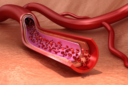
Blood is a liquid that keeps every human being alive. The blood vessels of an average adult male hold roughly 5.5 liters of the life-sustaining red fluid (Davy and Seals, 1994). The blood flow is essential for survival, and so is the infrastructure for its transport – the vasculature. This tissue consists of different cell types: smooth muscle cells, endothelial cells, cells from connective tissue, as well as an underestimated cell type that is gaining increasing attention: pericytes (Johnson et al., 2002).

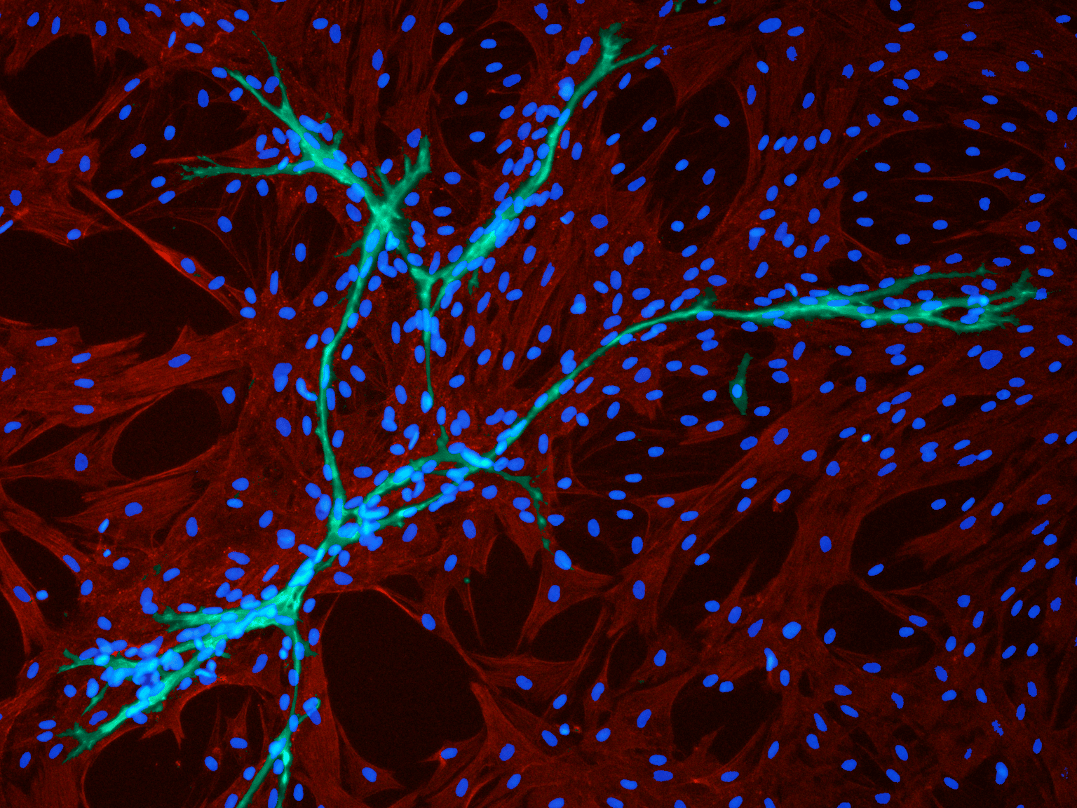
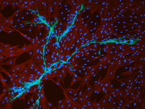
More than 140 years ago, Charles Marie Benjamin Rouget, a French physiologist, discovered pericytes, contractile cells that build a spider web around small vessels (Herndon et al., 2017). They are present in every vascularized tissue of the body, covering between 22 percent and 99 percent of the endothelial cell surface. Although scientists have known about pericytes for many years, they were not really sure of the role of these cells until recently. Even now, fundamental questions about where pericytes come from, what they do, and what they can become remain unanswered. However, new insights about cell surface markers, about interactions between pericytes and other cell types, and about their role in diseases are lifting the veil from their world (Bergers and Song, 2005).
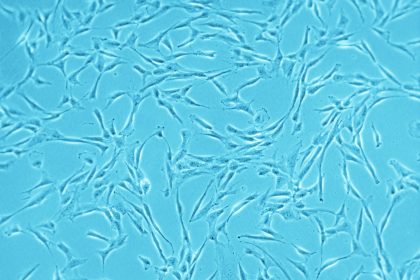
A particular challenge when working with pericytes is their heterogeneity. They display high variability in terms of ontogeny, localization and morphology. Moreover, some of them have stem cell properties, and can differentiate into adipocytes, osteoblasts, smooth muscle cells, chondroblasts and neural-like cells (Bouacida et al., 2012). In the past, many studies have focused on endothelial cells, and only in recent years has the importance of pericytes for the integrity of the vasculature been discovered. Pericytes are essential for maintaining a healthy vasculature. They work together with endothelial cells, building a functional unit and influencing each other in regulating microvascular stability, both through direct contact and through signaling molecules (Kelly-Goss et al., 2014). Pericytes also play important roles in regulating capillary blood flow, in angiogenesis, in hematopoiesis, in maintaining the permeability of the blood-brain barrier, in wound healing, and in leukocyte trafficking (Armulik et al., 2011).
Definition of pericytes – a heterogeneous population of stem cells
“It is very challenging to characterize the heterogeneity of vascular pericytes, which form a very dynamic population,” explains Dr. Frédéric Deschaseaux, leader of the group “Native MSC and Stemness” at the STROMALab in Toulouse, France, a research laboratory of the Etablissement Français du Sang. “It is hard to define good markers that can clearly identify all pericyte subpopulations.” Morphological features, such as the characteristic wrapping of cell processes around capillaries, can sometimes be misleading when using lower-resolution microscopy methods, as these do not allow a sure differentiation of pericytes from other perivascular cells.
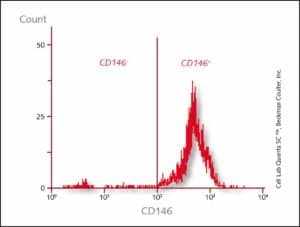
Also, there are no cell-type specific markers, and pericytes sometimes express the same surface proteins as other cells at the same anatomical location (Kelly-Goss et al., 2014). Commonly-used markers are SMA, Desmin, NG2 and PDGFR-β (Gerhardt and Betsholtz, 2003). “Different tissue locations can also have an impact on pericyte phenotype and function,” adds Deschaseaux. “To make the identification even more challenging, observations in vitro can be different from in vivo findings, as the medium used could have an impact on the phenotype and function of the cells. For example, the transcription factor TBX18 clearly defines pericytic cells in vivo, but not in vitro.”
The relationship between pericytes and mesenchymal stem cells (MSCs) has also been widely discussed. These two cell types reside in close proximity in the vascular wall and often express the same surface markers. Moreover, both have multipotential properties, and display regenerative capabilities. “The relationship between pericytes and MSCs is not really clear,” comments Deschaseaux. “In the lab, we have grown a population of pericytic progenitors, which can grow quickly, have an immature phenotype, and give rise to different mature populations. These cells are very close in phenotype and function to MSCs, but it is difficult to confirm whether they are MSCs, or subpopulations of MSCs.” (Stated by Bouacida et al., 2012). This is why the dynamics of the interactions between these two cell types remain difficult to describe clearly.
Dysfunctions lead to major pathological processes: pericytes in cancer
Pericytes are an integral part of the crosstalk among cells in the perivascular space. They interact not only with endothelial cells, but also with hematopoietic cells and immune cells, and are considered regulators of angiogenesis. Pericytes are important regulators of hematopoiesis, the mechanism that gives rise to all mature blood cells from hematopoietic stem cells (Itkin et al., 2016). When this endothelial balance is disrupted, pericyte dysfunction leads to pathological conditions affecting the vasculature, including diabetic retinopathy (Chou et al., 2014), fibrotic diseases (Rowley and Johnson, 2014), and cancer (Bergers and Song, 2005).
“Pericytes play a very important role in the tumor microenvironment, and in the expansion and dissemination of the tumor around the body,” says Deschaseaux. Due to their angiogenic potential, pericytes are essential during tumor neovascularization and for the stabilization of new blood vessels. Pericytes in the tumor microenvironment show different features than “normal” pericytes, suggesting the presence of an aberrant tumor-associated population, and a disturbed association with endothelial cells. New studies have shown a potential benefit in combining anti-cancer agents with anti-pericyte agents, as eliminating pericytes from the tumor-microenvironment severely impairs the blood supply of the tumor (Birbrair et al., 2014). Pericyte research is now focusing on identifying new drug targets in pericyte-associated pathological processes. A better understanding of pericyte biology, as well as insights into the molecular mechanisms by which pericytes and endothelial cells communicate, are prerequisites for the development of these new therapies.
A new tool in regenerative medicine
There is another side to pericytes. They are very important during tissue development, and in tissue regeneration processes, as they can be activated in response to tissue injury to contribute to the healing processes (van Dijk et al, 2015). “Pericytes play a strong role during tissue formation. When endothelial cells migrate into newly formed tissues during angiogenesis, pericyte populations lead the way to the top of the new vessels. These pericytes can then differentiate into mature cells of different specificities. When looking at bone injuries, or during bone development, vessels are formed by new cells in close interaction with pericytes, which express osteoblastic features,” explains Deschaseaux (Ono et al., 2014).
The regenerative potential of pericytes depends on the tissue they come from, and this characteristic has to be taken into account when designing pericyte-based regenerative strategies (Herrmann et al., 2016). “Pericytes show strong differentiation and proliferation potential in vitro. Both properties are very useful for cell therapy approaches. In addition, other perivascular cell types, adventitial cells, are described as “stem-like cells” exhibiting strong overlap on phenotype with pericytes (Hu and Xu, 2011). Therefore, efforts should continue that will enable scientists to clearly distinguish all different perivascular cells. New technologies such as single-cell RNA sequencing can provide data to better typify these cells, which will allow the development of highly precise regenerative therapies,” concludes Deschaseaux.