Welcome to PromoCell
We help scientists discover more every day by offering world-class cell culture products for research and industry applications that bridge the gap between discovery and clinical development.
About us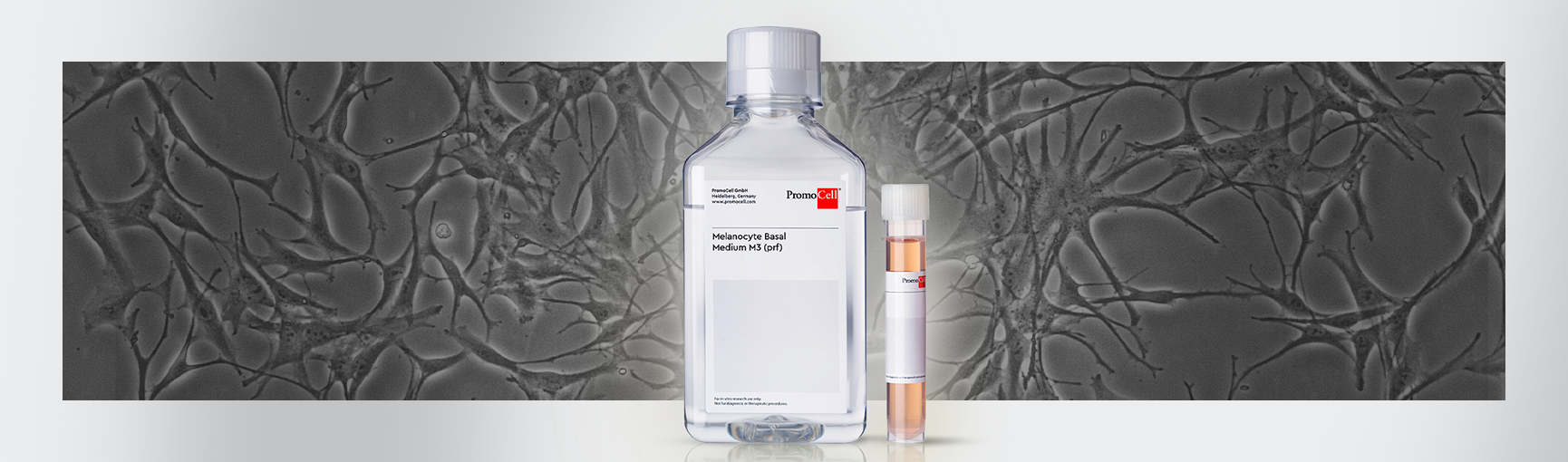


 and the SupplementMix tube.")
")
Find suitable products, scientific resources or answers to your questions within our technical library FAQ.