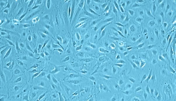
Human Dermal Microvascular Endothelial Cells (HDMEC)
 Powered by Bioz
Our products are available only through authorized distributors and resellers in your region.
Primary Human Dermal Microvascular Endothelial Cells (HDMEC) are isolated from the dermis of juvenile foreskin and adult skin (different locations). Since the dermis contains blood and lymphatic capillaries, HDMEC comprise Blood and Lymphatic Microvascular Endothelial Cells. Both have a common origin and can be identified by several markers.
In addition, HDMEC are available which are pre-screened for VEGF (vascular endothelial growth factor) response. VEGF is an important signaling protein involved in both vasculogenesis and angiogenesis. In vitro, VEGF has been shown to stimulate endothelial cell mitogenesis, cell migration, sprouting, and microvascular permeability.
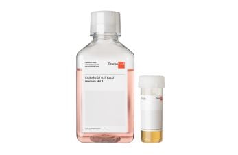
- Request our GMP grade cell culture media for endothelial cells.
- Our HDMEC (adult) are now also available from HLA-typed donors.
| Recommended plating density | 10,000 - 20,000 cells per cm2 |
| Passage after thawing | P2 |
| Tested markers | CD31 positive, Dil-Ac-LDL uptake positive |
| Guaranteed population doubling | > 15 |
| Recommended culture media* | C-22020,C-22022 |
*The catalog numbers in this table are for media in ready-to-use packaging.
PromoCell uses the Bioz AI engine to display scientific references for this product. This content is currently blocked because functional cookies are disabled.
Interested in our scientific references?
Click on the following link to load the content or enable functional cookies in the consent settings.
By clicking "Load content now", you agree to load content from Bioz, a third-party provider. This will set cookies on your device without changing your saved cookie preferences.