


Items 1-12 of 27 Results
-
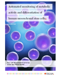
Automated monitoring of metabolic activity and differentiation of human mesenchymal stem cells
Detailed analysis of human stem cells offers the potential for the specific treatment of diseases by cell-based therapies. With an automated real-time analysis, we assess phenotypic changes during mesenchymal stem cell (MSC) differentiation. Application Notes
-
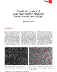
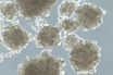
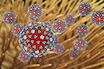
Standardized culture of assay-ready and fully functional human primary macrophages
Cryopreserved human macrophages are now available as a reliable source of standardized cells, and our ready-to-use format provides full experimental flexibility. With this easy-to-handle application, your macrophage research will be more efficient.
Application Notes
-
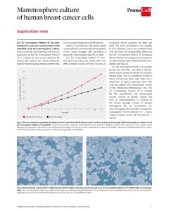
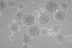
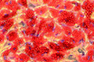
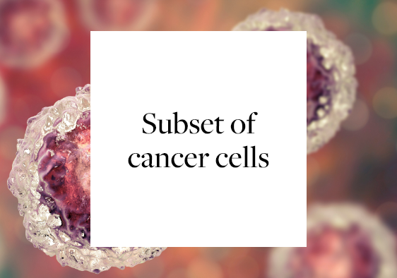
Mammosphere culture of human breast cancer cells
PromoCell 3D Tumorsphere Medium XF allows robust 3D culture even when using breast-cancer cell lines. Mammospheres are now regarded as highly valuable in vitro models for studies on tumor formation and metastasis.
Application Notes
-
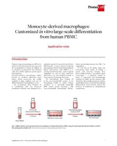
Monocyte-derived macrophages: Customized in vitro large-scale differentiation from human PBMC
Primary human macrophages are difficult to isolate and do not proliferate in culture. We offer a complete system for easy and cost-efficient generation of pure monocyte-derived macrophages.
Application Notes
-
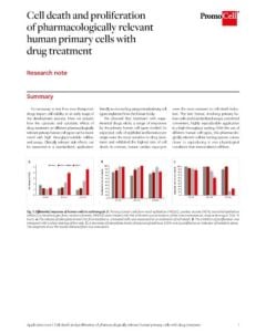
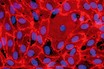
Cell death and proliferation of pharmacologically relevant human primary cells with drug treatment
Cytotoxic effects of a drug can be a crucial factor that differentiates a blockbuster from a failure. We assessed cytotoxic sensitivity of various primary cells in our portfolio using a high throughput-suitable assay.
Application Notes
-
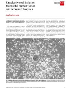
Unselective cell isolation from solid human tumor and xenograft biopsies
Do your primary cells from solid human tumors and xenografts need broad growth support? Then find out how to employ the serum-free, non-selective Cancer Cell Line Medium XF.
Application Notes
-
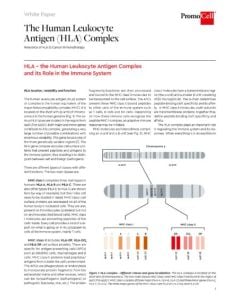
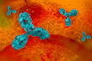
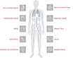
The human leukocyte antigen (HLA) complex
Learn more about HLA - The human leukocyte antigen complex and its role in the immune system.
White Papers
-
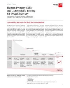
Human primary cells and cytotoxicity testing for drug discovery
Discover drug discovery cytotoxicity models for respiratory, cancer, cardiovascular, and dermatological research.
White Papers
-

Thawing frozen cells – detailed protocol for human primary cells
Thawing cells correctly is absolutely necessary when it comes to successfully starting reliable cell cultures. Find out how to thaw frozen primary cells properly in our detailed video protocol. Video
Video
-
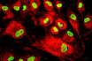
Cells in Action: Mitosis in Human Mesenchymal Stem Cells (MSCs)
Watch the process of cell division in PromoCell's human mesenchymal stem cells! These images were captured using Nanolive’s non-invasive technology, which allows us to access the dynamics of biological processes, such as mitosis, with unprecedented resolution.
Video
-
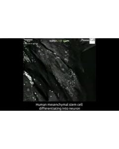
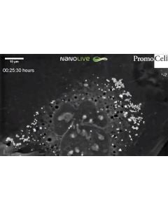
Cells in action: Differentiation of human mesenchymal stem cells into neurons
Cells in action: Differentiation of human mesenchymal stem cells into neurons In this video we can see the differentiation of PromoCell umbilical cord mesenchymal stem cells into neurons. Excitingly, to our knowledge this is one of the first high-resolution, long-term, live cell time-lapses showing this process. These cells were grown for 13 days in PromoCell complete mesenchymal stem cell neurogenic differentiation medium prior to capture of this film. Images were captured every 30 seconds over a 20-hour period using NanoLive’s 3D Cell Explorer.
Video
-
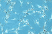
Cells in Action: Human Primary Keratinocytes at the stratum granulosum
Keratinocytes at the stratum granulosum contain two characteristic structures that are visible in this high resolution footage by Nanolive - the lamellar bodies and keratohyalin granules. While keratohyalin granules contain proteins involved in the aggregation of keratin filaments and in the formation of the cell envelope, lamellar bodies contain lipids and fuse with the plasma membrane in order to secrete the content into the extracellular space.
Video