3 Items
-

High-yield isolation of HUVECs in Endothelial Cell Growth Medium 2
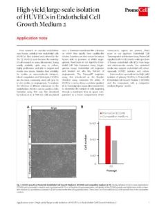
Here we show a procedure for a high-yield isolation of primary HUVECs in PromoCell’s Endothelial Cell Growth Medium 2 (ECGM2) and the comparison with a competitor medium. -

Cells in Action: membrane protrusions in HUVECs (Human Umbilical Vein Endothelial Cells)
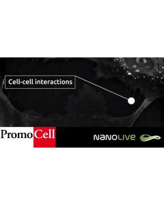
During angiogenesis, cell-matrix interactions are crucial. The cell matrix structures are involved in migration, invasion and survival of cells throughout the angiogenic process. In this video we can see filopodia and lamellipodia in great detail, thanks to Nanolive imaging's ability to view membrane deformations. These structures are essential for migration, cell-cell interactions, sensing of the cell environment and more. While filopodia appear as long, thin protrusion-like structures that emerge from the cellular membrane, lamellipodia are broad, sheet-shaped structures containing thin and short interconnected actin filaments. -

Cells in Action: Double mitosis of Human Umbilical Vein Endothelial Cells (HUVECs)
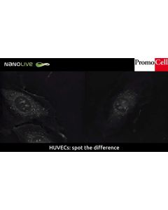
Images of cells undergoing mitosis are mind-blowing, and non more beautiful than those of our Human Umbilical Vein Endothelial Cells (HUVECs) captured by Nanolive's 3D Cell Explorer. On the right hand side, the cell seems to enter mitosis but the chromatids don't complete segregation and the cell returns to interphase without dividing. Exiting mitosis is controlled by proteolysis and cyclin dependent kinases (CDKs). Mitosis regulatory machinery sometimes detects errors and forces a return to interphase, as seen here. This kind of research helps develop understanding of the cell cycle, which can be used in myriad applications including cancer research.


