


RESOURCES
Items 25-36 of 144 Results
-
High-yield isolation of HUVECs in Endothelial Cell Growth Medium 2
Here we show a procedure for a high-yield isolation of primary HUVECs in PromoCell’s Endothelial Cell Growth Medium 2 (ECGM2) and the comparison with a competitor medium. Application notes
-
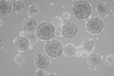
Epi-Fluorescence mitochondrial imaging in live single cells and tumorspheres
Mitochondria are the organelles that fuel life, which makes them extremely important for research. We offer a special dye for live-cell and fixed-cell imaging. Use this dye to boost your mitochondria research. Find out how.
Application notes
-
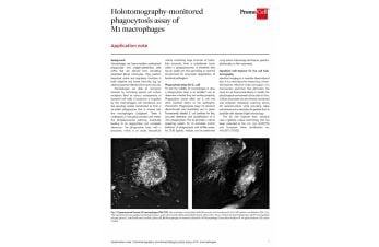
Holotomography-monitored phagocytosis assay of M1 macrophages
Phagocytosis is an essential process for pathogen clearance and is the subject of research for many immunologists. Find out more about PromoCell's phagocytosis assay for fluorescence microscopy and how you can implement this method in your lab.
Application notes
-
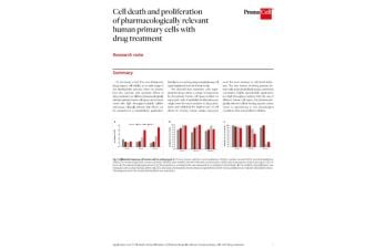
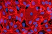
Cell death and proliferation of pharmacologically relevant human primary cells with drug treatment
Cytotoxic effects of a drug can be a crucial factor that differentiates a blockbuster from a failure. We assessed cytotoxic sensitivity of various primary cells in our portfolio using a high throughput-suitable assay.
Application notes
-
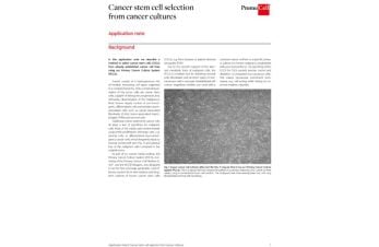
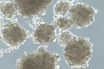
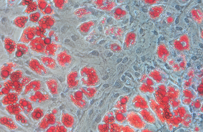
Cancer stem cell selection from cancer cultures
Traditional culture systems for cancer cells all share a lack of specific growth support for malignant cells. PromoCell's Primary Cancer Culture System is a reliable tool for depleting stromal cells, fibroblasts, and all other types of non-cancerous cells.
Application notes
-
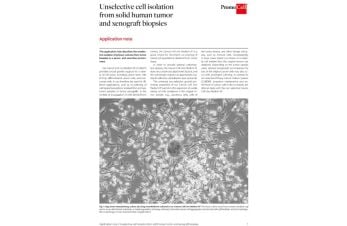
Unselective cell isolation from solid human tumor and xenograft biopsies
Do your primary cells from solid human tumors and xenografts need broad growth support? Then find out how to employ the serum-free, non-selective Cancer Cell Line Medium XF.
Application notes
-
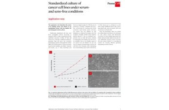
Standardized culture of cancer cell lines under serum and xeno-free conditions
Undefined media supplements have unwanted effects on cell cultures. Learn how to obtain more accurate and comparable results with PromoCell’s Cancer Cell Line Medium XF.
Application notes
-
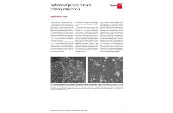
Isolation of patient-derived primary cancer cells
Isolate tumor cells from patient samples and transform them into long-term cell cultures. Learn more about the primary cancer culture system and how it compares to other established culture methods.
Application notes
-
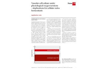
Vascular cell culture under physiological oxygen tensions – implications for cellular redox homeostasis
If the amount of reactive oxygen species exceeds the cells antioxidant capacity, your research subjects are stressed. Find out how you can shift your cell culture conditions and establish physiological conditions.
Application notes
-
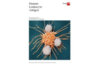
The importance of HLA for the development of cell therapies
Download our white paper to understand the role Human Leukocyte Antigen plays in the development of cell therapies.
White papers
-
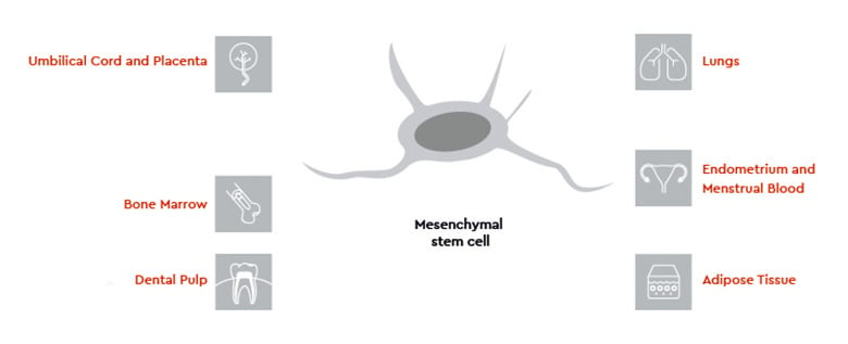
Mesenchymal stem cells: why optimizing manufacturing processes is key for a successful application
Discover the reasons why an optimized manufacturing process is essential for successful application of mesenchymal stem cells.
White papers
-
How to incorporate organoid cultures into your research – expert interview
Our organoid expert Dr. Elfie Rödel shares her top tips for working with 3D organoid cultures into research.
White papers