3D cell culture
Find suitable products, scientific resources or answers to your questions within our technical library FAQ.
Checkout using your account
This form is protected by reCAPTCHA - the Google Privacy Policy and Terms of Service apply.
Checkout as a new customer
Creating an account has many benefits:
Reliable expansion of CD34⁺ hematopoietic stem and progenitor cells (HSPCs) in in vitro culture is essential when you’re developing cell and gene therapy applications.
CD34⁺ HSPCs generate all mature blood and immune cells, making them an important tool for cell and gene therapy. Hematopoietic progenitor cells (HPCs), a key HSPC subset, drive downstream differentiation. Expanding these cells outside the body can be challenging. Donor variability, undefined serum components, and differentiation pressure during culture can make it challenging to reliably expand HSPCs in vitro.
You can isolate CD34⁺ HSPCs from mobilized peripheral blood, bone marrow, and cord blood. Each source has a different frequency and expansion potential of CD34⁺ cells, making baseline cell characterization essential.1–3
A typical isolation workflow includes:
Standardizing these steps early reduces variability in downstream expansion.
Native cell sources rarely yield enough CD34⁺ HSPCs for therapeutic or research use. In vitro cell expansion helps you overcome this bottleneck. Expansion helps you increase the number of CD34+ HSPCs without driving differentiation.
To expand CD34⁺ HSPCs, seed cells at a defined density and culture them with cytokines that promote proliferation.6 TPO, SCF, FLT3 ligand, and IL 3 are commonly used to support the expansion of HPCs. CD34+ HSPC expansion typically requires frequent medium changes and fed-batch culture.7 These approaches help reduce the accumulation of inhibitory factors secreted by differentiating cells.
Looking for a defined medium optimized for HPC expansion?
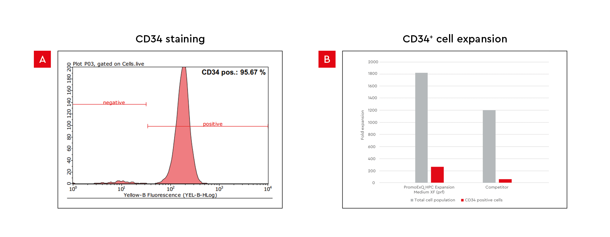
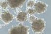
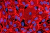
Figure 1: CD34⁺ HPC expansion in culture. A) The figure shows flow cytometry analysis of our CD34⁺ progenitor cells on day 0 of culture. B) Representative data of the expansion of human cord blood CD34⁺ cells after 13 days of culture. Cells were expanded using the HPC Expansion Medium XF or competitor medium.
Each variable in your HSPC expansion protocol can influence the quality of your CD34⁺ cells. The table below summarizes the variables that determine expansion quality, why each one matters, and what to measure or record as evidence.
| Variable | Impact on CD34⁺ expansion | Recommended action |
|---|---|---|
| Starting cell source and donor variability8,9 | Affects expansion potential and baseline phenotype | Record donor/source; set acceptance criteria for viability and %CD34 pre-culture |
| Medium format (serum-containing vs serum-free [SF]/xeno-free [XF]) | Undefined serum components can increase variability and differentiation pressure | Use SF/XF where possible; document lot strategy and comparability readouts |
| Cytokine supplementation strategy9 | Controls proliferation vs differentiation balance | Define cytokine mix and timing; track fold expansion and %CD34 retention |
| Culture density and duration6 | Impacts nutrient availability and lineage skew | Standardize seeding density; track viability, %CD34, and output cell counts at fixed timepoints |
| Functional output (e.g., Colony-Forming Unit [CFU] assay)10 | Provides evidence of progenitor proliferation and lineage potential | Run CFU as a periodic check; record colony types/counts using consistent scoring rules |
Table 1: Variables influencing HSPC expansion quality.
These variables shape the functional composition of the expanded population, including the proportion and performance of HPCs required for downstream applications. Control them from the start to avoid compatibility issues, especially if you change medium format mid-study.
Serum introduces undefined variables into your HSPC culture. Growth factor concentrations, lipid profiles, and contaminants vary between lots, and that variability affects your downstream results.11
Defined, serum-free media overcome these variations, offering several advantages over serum-containing media:
The bar in CD34⁺ HSPC expansion for therapy development is much higher than that for research. Compared to research applications, therapeutic use requires:
CD34⁺ HSPC differentiation enables multiple downstream applications, including production of the following:
Getting the most from these applications means building an expansion workflow that is scalable, from early research through to further manufacturing.
There are three stages to moving CD34⁺ HSPC expansion from a research setting toward a more regulated environment.
Establish baseline expansion performance using your cell source of choice.
Use defined media solutions to improve consistency and reduce variability.
Align your workflow with regulatory expectations before scale-up.
*“GMP-grade” is a branding term used by PromoCell to denote reagents that are manufactured at the PromoCell manufacturing facility in Heidelberg, Germany, under strictly controlled processes to meet stringent product specifications and customer requirements. Reagents manufactured at PromoCell are produced according to EXCiPACT™ GMP standards, a quality management system that builds on our ISO 9001:2015 certification. Risk assessment procedures are carried out at the customer site.
Ready to optimize your HSPC expansion? Improve expansion consistency, support phenotype retention, and reduce variability with media solutions tailored to your application and cell source.
References
Manufactured in compliance with the EXCiPACT™ GMP certification standard.
Defined, animal-component free, and protein-free cryopreservation medium for optimal cell storage.
Serum-free and xeno-free medium for expansion of primitive human hematopoietic cells.
Related products
 Product
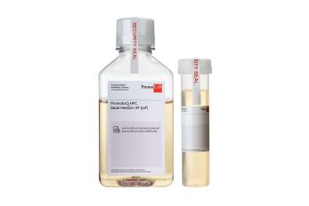
PromoExQ HPC Expansion Medium XF
Product
PromoExQ HPC Expansion Medium XF
 Product
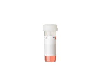
Freezing Medium Cryo-SFM Plus
Product
Freezing Medium Cryo-SFM Plus
 Product
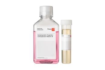
Hematopoietic Progenitor Cell Expansion Medium XF
Product
Hematopoietic Progenitor Cell Expansion Medium XF
Have a look at other Research Areas
PromoCell uses HubSpot to provide LiveChat support. This feature is currently blocked due to your cookie preferences.
Accept cookies to enable LiveChat