


Citations
PromoCell uses the Bioz AI engine to display product citations. This content is currently blocked due to your cookie preferences.
Normal Human Epidermal Keratinocytes (NHEK) GM3
Primary Human Keratinocytes isolated in serum-free and BPE-free Keratinocyte Growth Medium 3. Available from juvenile foreskin or adult skin from single or pooled donors.
Normal Human Epidermal Keratinocytes (NHEK) adult, isolated in Growth Medium 3, single donor
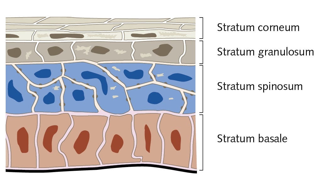
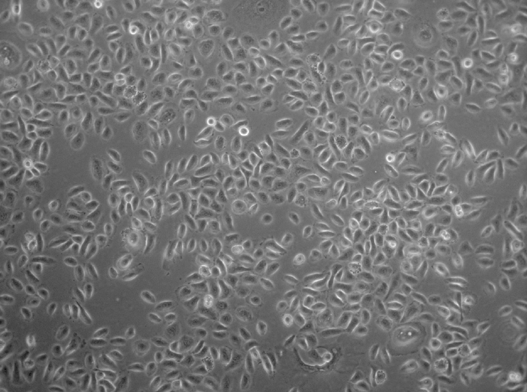
Primary Normal Human Epidermal Keratinocytes (NHEK) GM3 are available from single or from pooled donors isolated from the epidermis of juvenile foreskin or adult skin from different locations like the face, the breasts, the abdomen, and the thighs. They are the major cell type in the epidermis, making up about 90% of the cells.
Epidermal keratinocytes originate in the stratum basale and move up through the layers of the epidermis. During this movement, they undergo gradual differentiation and morphology changes until they reach the stratum corneum, where they form a layer of nucleus-free, flat, and highly keratinized squamous cells. This layer forms an effective barrier to the entry of infectious agents into the body and minimizes moisture loss.
Keratinocytes are also able to produce a variety of cytokines, growth factors, interleukins and complement factors. Therefore keratinocytes are important for wound healing, inflammation, and immune response.
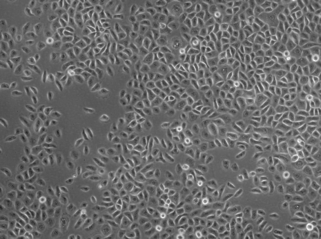
Our Primary Normal Human Epidermal Keratinocytes GM3 are isolated using the improved serum-free and BPE-free Keratinocyte Growth Medium 3. The optimized formulation supports a growth of fast proliferating and homogenous cell population without the need for extracellular matrix coatings or feeder cells.
Normal Human Dermal Fibroblasts (NHDF) or Normal Human Epidermal Melanocytes M3 (NHEM M3) from the same donor are available on request.
Our NHEK are now also available from HLA-typed donors.
Recommended plating density | 5000 cells per cm2 |
Passage after thawing | P2 |
Tested markers | Cytokeratin positive |
Guaranteed population doubling | > 15 |


We use a classification system similar but not identical to the Fitzpatrick Skin Classification. The Fitzpatrick classification has six different categories (phototypes I-VI) which correlate with the level of skin pigmentation (melanin) and sunburn following sun exposure. Fitzpatrick I corresponds with the lightest of skin complexions, while Fitzpatrick VI corresponds with the darkest skin.
- I: Pale white skin, blue/hazel eyes, blond/red hair, always burns, does not tan
- II: Fair skin, blue eyes, burns easily, tans poorly
- III: Darker white skin, tans after initial burn
- IV: Light brown skin, burns minimally, tans easily
- V: Brown skin, rarely burns, tans darkly easily
- VI: Dark brown or black skin, never burns, always tans darkly
At PromoCell, we have knowledge of the patients’ skin color (white, brown or black skin), color of eyes and hair, but we don't have any details about the burning/tanning abilities. We therefore classify our tissue donors as follows:
- Light (comprising phototypes I and II)
- Moderate (comprising phototypes III and IV)
- Dark (comprising phototypes V and VI)
Information on the phototype is available for most cell lots isolated from juvenile or adult skin.
Short protocol:
- Trypsinize the cells as usual
- Centrifuge and resuspend in suitable cold freezing medium at a density of 1-4 x 10⁶ cells/ml
- Cool down the cells slowly to -80°C (approx. -1°C per min). We recommend to use "Mr. Frosty" from Nalge or "CoolCell" from Biocision; which both provide gradual and controlled cooling rates when placed in a -80°C freezer overnight.
- Transfer the vials into liquid nitrogen for long-term storage
- Mesenchymal Stem Cells (C-12974/C-12971/C-12977) need Fibronectin-coating when grown in PromoCell MSC Growth Medium XF (C-28019) and when differentiated in MSC Neurogenic (C-28015); Adipogenic (C-28016); or Osteogenic (C-28013) Differentiation Media.
- Human monocyte-derived macrophages (C-12914/C-12916/C-12915/C-12917) must be seeded into Fibronectin-coated culture vessels in combination with PromoCell's M1- and M2-Generation Media XF (C-28055; C-28056).
- For efficient induction of osteoblast mineralization with PromoCell's Osteoblast Mineralization Medium (C-27020); the TC plates should be pre-coated with collagen type I.
- Liquid phase storage provides a consistent temperature of -196°C; a longer holding time and a greater vial capacity but involves the risk of contamination issues.
- Storage in the gas phase is very safe with respect to contaminations but the holding time of the cells is shorter and the vial capacity is reduced.
General protocol for recovery of anchorage-dependent primary cells: Remove vial from liquid nitrogen; transport it on dry ice to the cell culture lab. Thaw in a 37°C waterbath for approx. 2 min; until it is just defrosted. Keep the vial immersed in water until just below the screw cap during thawing and only remove it shortly after approx. 90 sec to check the progress. Do not repeatedly insert and remove the vial while thawing in water. Carefully disinfect the vial with plenty of 70% EtOH under the laminar flow hood and aseptically transfer the thawed suspension into an appropriate TC dish with growth medium (pre-warmed in the incubator for > 30 min). The cells usually attach within a few hours. Perform media change after 24 hrs at the latest to remove residual DMSO from the freezing media. Additional information can be found in the instruction manuals of our cells.
The optimal calcium concentration for both proliferation and differentiation of keratinocytes depends on the species and also on the media formulation. To keep primary human keratinocytes in the proliferative status concentrations between 0.03 and 0.15 mM (PromoCell Keratinocyte Growth Medium 2: 0.06 mM) are generally used. Increasing the calcium above 1 mM will induce terminal differentiation and lead to the loss of proliferative activity.


PromoCell uses the Bioz AI engine to display scientific citations for this product. This content is currently blocked because you haven’t enabled functional cookies.
To see this content, please click the cookie settings icon (bottom left corner of the screen) and enable functional cookies.
Alternatively, you can load the content directly:
By clicking "Load content now", you agree to load content from Bioz, a third-party provider. This sets cookies on your device, without changing your saved cookie preferences.
Recommended products

Keratinocyte Growth Medium 3
Serum-free and BPE-free cell culture medium for the cultivation of human keratinocytes from the epidermis of juvenile foreskin or adult skin.