Keratinocytes: What you should know
Keratinocytes are the predominant cell type of the epidermis, the outermost layer of the skin. They form and maintain the epidermal barrier, help protect the body from external substances, and contribute to wound healing, inflammation, pigmentation biology, and skin model research.
Because keratinocytes are central to epidermal structure and function, primary human keratinocytes are widely used in dermatology, toxicology, cosmetic testing, wound-healing research, and 3D skin model development.
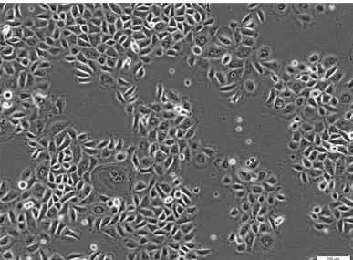
What are keratinocytes?
Keratinocytes are specialized epithelial cells that make up most of the epidermis. They are named after keratin, a structural protein that contributes to the strength and protective function of the skin.
In the epidermis, keratinocytes form tightly connected cell layers. These layers help create a physical barrier that protects the body from infectious agents, environmental stressors, and excessive water loss. Keratinocytes also produce cytokines, growth factors, interleukins, and complement factors, which allows them to participate in immune and inflammatory responses.
Where are keratinocytes found in the skin?
Keratinocytes are found in the epidermis. They originate in the stratum basale, the deepest layer of the epidermis, and gradually move upward through the epidermal layers as they differentiate.
During this process, keratinocytes pass through the stratum spinosum and stratum granulosum before reaching the stratum corneum. In the stratum corneum, they are present as flattened, nucleus-free, highly keratinized cells that contribute to the final barrier layer of the skin.1
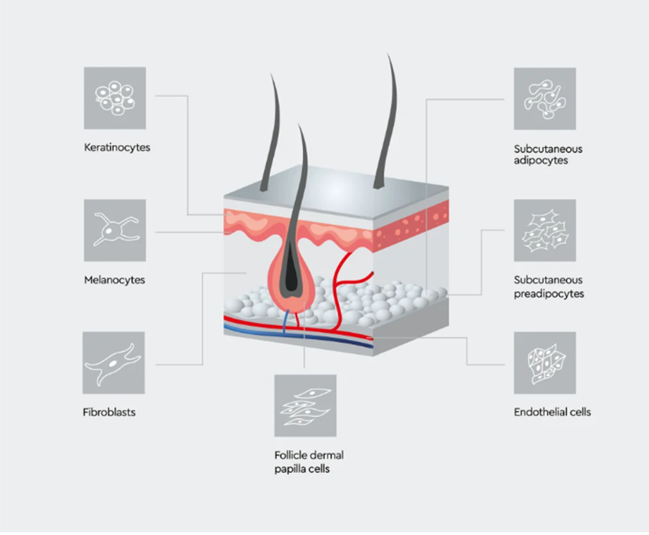
Figure 1: Skin-associated cell types relevant to in vitro skin research.
Keratinocytes interact with other skin-associated cells, including melanocytes, fibroblasts, endothelial cells, adipocytes, preadipocytes, and follicle dermal papilla cells, depending on the biological process or model system being studied.
How do keratinocytes differentiate?
Keratinocyte differentiation is the process by which proliferating basal keratinocytes mature into the flattened, keratin-rich cells of the stratum corneum.
Keratinocytes proliferate in the basal layer of the epidermis. As they move toward the skin surface, they undergo gradual changes in morphology, protein expression, and function. They begin to produce structural proteins and differentiation-associated markers that support epidermal organization and barrier formation.
This differentiation process is regulated by multiple biological factors, including extracellular signals, calcium concentration, and epigenetic mechanisms. A balanced differentiation process is essential for normal epidermal renewal and skin barrier function.2
What do keratinocytes do?
Keratinocytes help form the skin barrier, maintain epidermal structure, and participate in immune and wound-healing responses.
| Function | Why it is important |
|---|---|
| Barrier formation | Helps protect the body from external substances and infectious agents |
| Moisture retention | Helps reduce water loss through the skin |
| Structural support | Maintains the organization and stability of the epidermis |
| Immune signaling | Supports communication with immune cells after barrier disruption or injury |
| Tissue repair | Contributes to re-epithelialization during wound healing |
Table 1: Key functions of keratinocytes
Overview of the main biological functions of keratinocytes in epidermal barrier formation, immune signaling, and tissue repair.
Keratinocytes are therefore more than passive barrier cells. They actively sense changes in their environment and help coordinate skin responses to injury, inflammation, and external stress.
Keratinocytes cell culture
How are keratinocytes cultured?
Human keratinocytes can be cultured in two-dimensional monolayer systems and in more complex three-dimensional skin models.3 Normal Human Epidermal Keratinocytes (NHEK) are commonly used for in vitro studies of epidermal biology, differentiation, barrier formation, wound healing, and inflammatory responses.
Normal Human Epidermal Keratinocytes (NHEK) can be isolated from juvenile foreskin or adult skin from different locations, including the face, breast, abdomen, and thighs. Depending on the research question, keratinocytes from single donors or pooled donors may be used.
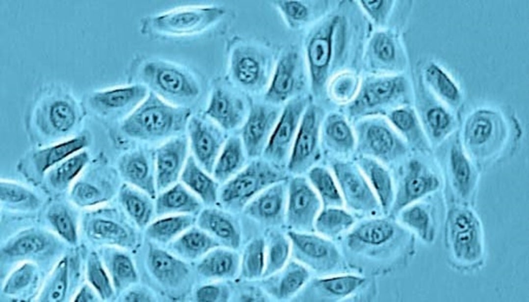
Figure 2: Normal Human Epidermal Keratinocyte culture in phase contrast.
NHEK show the typical epithelial morphology used to assess culture appearance.
Culture conditions are important because keratinocyte proliferation and differentiation are sensitive to the culture environment. Medium composition, donor source, passage number, calcium concentration, and antibiotic use can all influence cell behaviour.
For practical data on keratinocyte morphology and growth performance in different culture conditions, see our application note on isolating fast-proliferating and homogeneous keratinocyte populations.
For keratinocyte culture, Keratinocyte Growth Medium 2 provides a serum-free medium option for epidermal keratinocytes. Keratinocyte Growth Medium 3 may be considered where a serum-free and BPE-free medium is preferred. For an overview of related products, see primary keratinocytes and culture media.
How are keratinocytes characterized?
Keratinocytes are characterized using morphology, growth behaviour, and marker expression. In culture, normal human keratinocytes typically show a cobblestone-like morphology.
Keratinocyte marker expression changes during differentiation. At different differentiation stages, keratinocytes express specific keratins and additional markers associated with epidermal maturation and barrier formation.
| Marker | Relevance |
|---|---|
| Cytokeratins | Common epithelial and keratinocyte-associated markers |
| Involucrin | Marker associated with keratinocyte differentiation |
| Loricrin | Marker of late differentiation and cornified envelope formation |
| Transglutaminase | Supports cornified envelope formation |
| Filaggrin | Associated with epidermal barrier formation |
| Caspase-14 | Associated with terminal keratinocyte differentiation |
Table 2: Common keratinocyte characterization markers
Summary of markers commonly used to assess keratinocyte identity, differentiation, and barrier associated maturation.
Marker selection depends on the experimental model, differentiation stage, and research question. Combining marker analysis with morphology and growth performance can help confirm the suitability of the culture for a specific application.
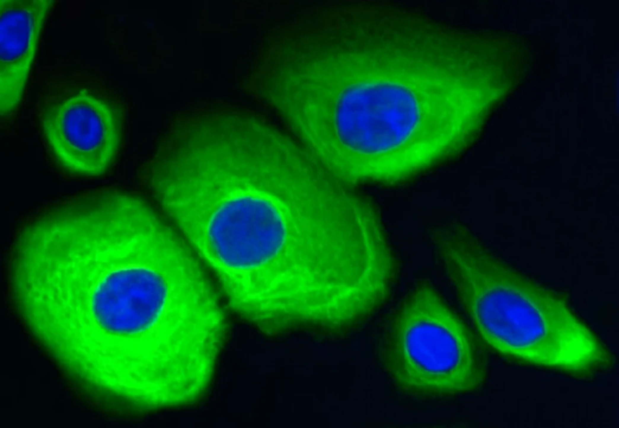
Figure 3: Cytokeratin staining of Normal Human Epidermal Keratinocytes.
NHEK stained for cytokeratin, with nuclei counterstained with DAPI.Normal Human Epidermal Keratinocyte culture in phase contrast.
How do keratinocytes interact with other skin cells?
Keratinocytes interact with several other skin cell types, including fibroblasts, melanocytes, Langerhans cells, lymphocytes, and other immune cells. These interactions help maintain skin homeostasis and support coordinated responses to injury and inflammation.
Keratinocytes and fibroblasts
Keratinocyte-fibroblast communication is important for skin homeostasis and wound healing.4 Fibroblasts are located in the dermis and contribute to extracellular matrix production, tissue structure, and repair processes. Keratinocytes and fibroblasts communicate through paracrine signaling, and disruption of this cross-talk can contribute to impaired wound healing and chronic wounds.
For related skin repair models, see Normal Human Dermal Fibroblasts (NHDF) and the application note on a 3D in vitro wound healing model.
Keratinocytes and melanocytes
Keratinocytes also interact closely with melanocytes. Melanocytes produce melanin, which helps protect keratinocytes from UV-induced DNA damage. Keratinocytes, in turn, influence melanocyte proliferation, differentiation, and melanogenesis.5
This interaction is relevant for research into pigmentation, UV response, epidermal homeostasis, and skin disease. For related products, see Normal Human Epidermal Melanocytes (NHEM).
Keratinocytes and immune cells
Keratinocytes contribute to immune communication in the skin. After barrier disruption or pathogen exposure, keratinocytes can produce cytokines and other signaling molecules that influence immune cell recruitment and activation.
Through these functions, keratinocytes help connect the physical barrier of the epidermis with the immune response of the skin.
What role do keratinocytes play in wound healing and inflammation?
Keratinocytes are essential for re-epithelialization, the process by which the epidermis is restored after injury.6
After skin damage, keratinocytes become activated, migrate toward the wound area, and proliferate to help close the defect. During this process, keratinocytes interact with fibroblasts and immune cells to support tissue repair. Impaired keratinocyte function can contribute to delayed wound closure and chronic wounds.
Keratinocytes are also involved in inflammatory skin responses. When the epidermal barrier is breached or pathogens enter the skin, keratinocytes can produce cytokines that send activating or regulatory signals to immune cells.7 This makes them relevant for research into allergic skin diseases and chronic inflammatory conditions such as psoriasis.8
What are keratinocytes used for in research?
Keratinocytes are used in research areas where epidermal biology, skin barrier function, cell differentiation, wound healing, inflammation, or skin responses to compounds are relevant.
| Research area | How keratinocytes are used |
|---|---|
| Epidermal biology | Study epidermal development, renewal, and differentiation |
| Wound healing | Investigate keratinocyte migration, proliferation, and re-epithelialization |
| Inflammation research | Study cytokine signaling and immune interactions in the skin |
| Toxicology | Assess skin responses to test compounds |
| Cosmetic testing | Support in vitro evaluation of skin-relevant effects |
| Skin aging research | Investigate changes in epidermal function and repair |
| Cancer research | Study mechanisms relevant to epithelial transformation and skin cancer |
| 3D skin models | Build epidermal or full-thickness skin model systems |
Table 3: Keratinocyte applications in research
Overview of research areas where keratinocytes are used to study skin biology, repair, inflammation, toxicology, and 3D skin models.
For additional application context, explore how primary human skin cells are used in primary cell culture in dermatology and how keratinocyte-based systems contribute to 3D skin models in dermatology research.
Keratinocytes by PromoCell
PromoCell offers [Normal Human Epidermal Keratinocytes (NHEK)] from single or pooled donors. Cells are isolated from juvenile foreskin or adult skin from different anatomical locations, including the face, breast, abdomen, and thighs.
Additional donor information, such as age, sex, and skin pigmentation, is available for each produced lot. Each lot is tested for cell type-specific markers, growth performance, and the absence of relevant viral and microbial contaminants.
To support keratinocyte culture workflows, PromoCell also offers dedicated keratinocyte media, including [Keratinocyte Growth Medium 2 and Keratinocyte Growth Medium 3. Related skin cell products, such as Normal Human Dermal Fibroblasts (NHDF) and Normal Human Epidermal Melanocytes (NHEM), can support studies of skin cell cross-talk, pigmentation biology, and wound-healing models.
Explore our keratinocyte portfolio to find primary human keratinocytes, optimized culture media, and related resources for skin cell research.
Stay informed about skin cell research Working with keratinocytes, fibroblasts, melanocytes, or 3D skin models? Explore our dermatology and skin cell culture resources to learn more about primary human skin cells, culture conditions, and application-relevant models.
FAQs about keratinocytes
References
Expand
- Eckert RL, Rorke EA. Molecular biology of keratinocyte differentiation. Environ Health Perspect. 1989 Mar;80:109–116. doi:10.1289/ehp.8980109
- Lee J-H. Keratinocyte Differentiation and Epigenetics. In: Lu Q, Chang CC, Richardson BC, editors. Epigenetics and Dermatology. Academic Press; 2015. p. 37–52. doi:10.1016/B978-0-12-800957-4.00003-5
- Rasmussen C, Thomas‑Virnig C, Allen‑Hoffmann BL. Classical human epidermal keratinocyte cell culture. Methods Mol Biol. 2013;945:161–175. doi:10.1007/978‑1‑62703‑125‑7_11
- Wojtowicz AM, Oliveira S, Carlson MW, Zawadzka A, Rousseau CF, Baksh D. The importance of both fibroblasts and keratinocytes in a bilayered living cellular construct used in wound healing. Wound Repair Regen. 2014 Mar‑Apr;22(2):246–255. doi:10.1111/wrr.12154
- Hirobe T. Keratinocytes regulate the function of melanocytes. Dermatologica Sinica. 2014;32(4):200–204. doi:10.1016/j.dsi.2014.05.002
- Pastar I, Stojadinovic O, Yin NC, Ramirez H, Nusbaum AG, Sawaya A, Patel SB, Khalid L, Isseroff RR, Tomic‑Canic M. Epithelialization in wound healing: a comprehensive review. Adv Wound Care (New Rochelle). 2014 Jul 1;3(7):445–464. doi:10.1089/wound.2013.0473
- Albanesi C, Scarponi C, Giustizieri ML, Girolomoni G. *Keratinocytes in inflammatory skin diseases*. Curr Drug Targets Inflamm Allergy. 2005 Jun;4(3):329–334. doi:10.2174/1568010054022033
- Albanesi C, Madonna S, Gisondi P, Girolomoni G. The interplay between keratinocytes and immune cells in the pathogenesis of psoriasis. Front Immunol. 2018;9:1549. doi:10.3389/fimmu.2018.01549
Primary Human Epidermal Melanocytes isolated in PMA-containing Melanocyte Growth Medium from the epidermis of juvenile foreskin.
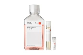
Serum-free and BPE-free cell culture medium for the cultivation of human keratinocytes from the epidermis of juvenile foreskin or adult skin.
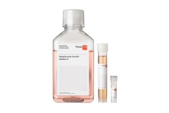
Serum-free cell culture medium for keratinocytes from the epidermis.
Primary Human Keratinocytes isolated from the epidermis of juvenile foreskin or adult skin from single or pooled donors.
Related products