Our understanding of the respiratory system has improved drastically over the last few years. In vitro lung models have undoubtedly played an instrumental role in enhancing our understanding of lung diseases and in finding new, potentially life-saving therapies. These models are invaluable tools in lung biology studies, respiratory disease research, drug discovery and testing, environmental exposure studies, and personalized medicine approaches.1 In this article, we explore how innovative in vitro lung models are transforming respiratory research and drug development.
What are in vitro lung models?
In vitro lung models are advanced cell culture systems used in the laboratory or clinic to replicate the physiological and pathological aspects of the human lung outside the body. These models provide a controlled environment for studying lung biology and disease mechanisms and for testing potential treatments.2 By closely mimicking the complex interactions within the human lung, in vitro lung models offer researchers a powerful tool for gaining insights that were once challenging to obtain.The mechanics behind in vitro lung models
The lung, the most important organ in the human respiratory system, primarily comprise alveolar epithelial cells (type I and type II cells), pulmonary fibroblasts, bronchial epithelial cells, and the small airway epithelium (basal, intermediate, ciliated, mucin-producing, and club cells) (Figure 1).3 In vitro respiratory models incorporate various types of lung cells (e.g., bronchial epithelial cells, pulmonary fibroblasts, and small airway epithelial cells) and extracellular matrix (ECM) components to recapitulate some aspects of the cellular diversity and physiology of the human lung.4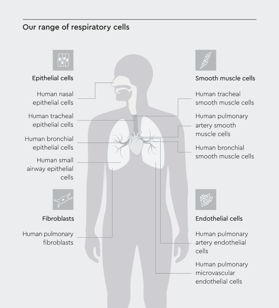
Figure 1: Cell composition of the human respiratory tract.
To help you establish in vitro lung models, we offer a broad range of human respiratory cell types, including epithelial cells, endothelial cells, fibroblasts, and smooth muscle cells.
Different types of in vitro lung models
Over the years, researchers have developed various in vitro lung models (Figure 2). From traditional two-dimensional (2D) cell cultures to advanced 3D organoids and organotypic models, each offers unique advantages. In vitro respiratory models include:- 2D culture models: These are the simplest form of in vitro lung models, where cells are grown as monolayers on plastic or glass.5 They are easy to set up and allow for fundamental cell and molecular biology studies and high-throughput screening. However, they lack the complex 3D structure of the lungs, which can limit their physiological relevance.
- Air-liquid interface (ALI): ALI culture systems may help bridge the gap between 2D and 3D, as they can be used to generate stable and functional 3D human airway models incorporating primary human cells through an intermediate 2D cell expansion phase. For additional details on setting up ALI culture systems, we suggest our AppNote Air-liquid interface culture system for standardized respiratory research.
- 3D organoids: When cells are cultured in extracellular matrix (ECM) gels or scaffolds, cells may self-organize into simple organ-like structures.5 3D organoids are more complex than 2D models and can better mimic the in vivo environment, enabling analysis of cell crosstalk, differentiation, and infection. However, they can be challenging to set up and maintain.
- Organotypic models: These models mimic the realistic multicellular architecture of the lung and provide a more accurate representation of the in vivo environment. Cells arranged on permeable supports to form polarized air-liquid interface cultures recreate airway and alveolar architecture that allow for advanced experimentation.5 They can incorporate multiple cell types and may be used to study cell-cell and cell-matrix interactions. However, they are the most complex and challenging to set up.
- Microfluidic “lung-on-chip” devices: These devices incorporate engineered microchannels and membranes to enable the application of airflow, liquid perfusion, and mechanical strain to mimic the dynamic lung microenvironment.6
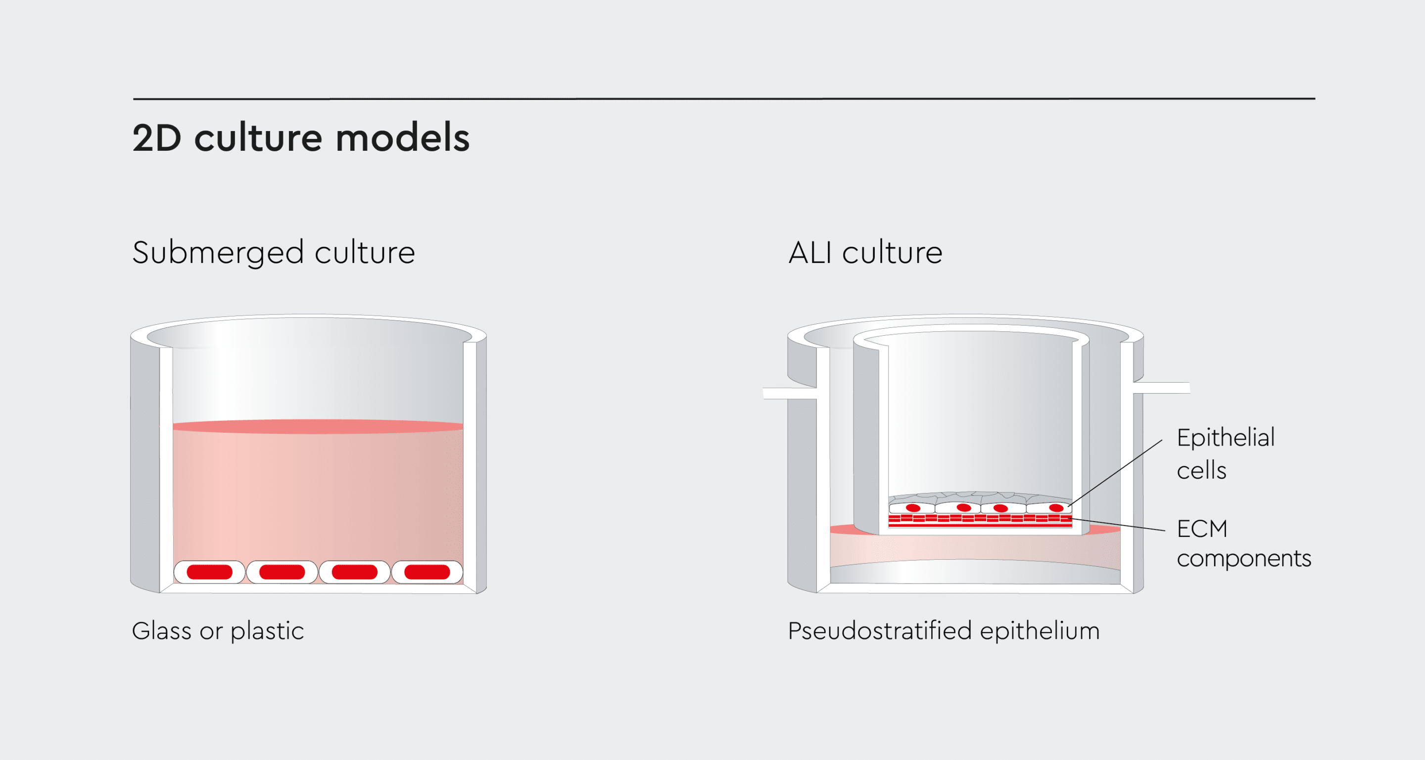
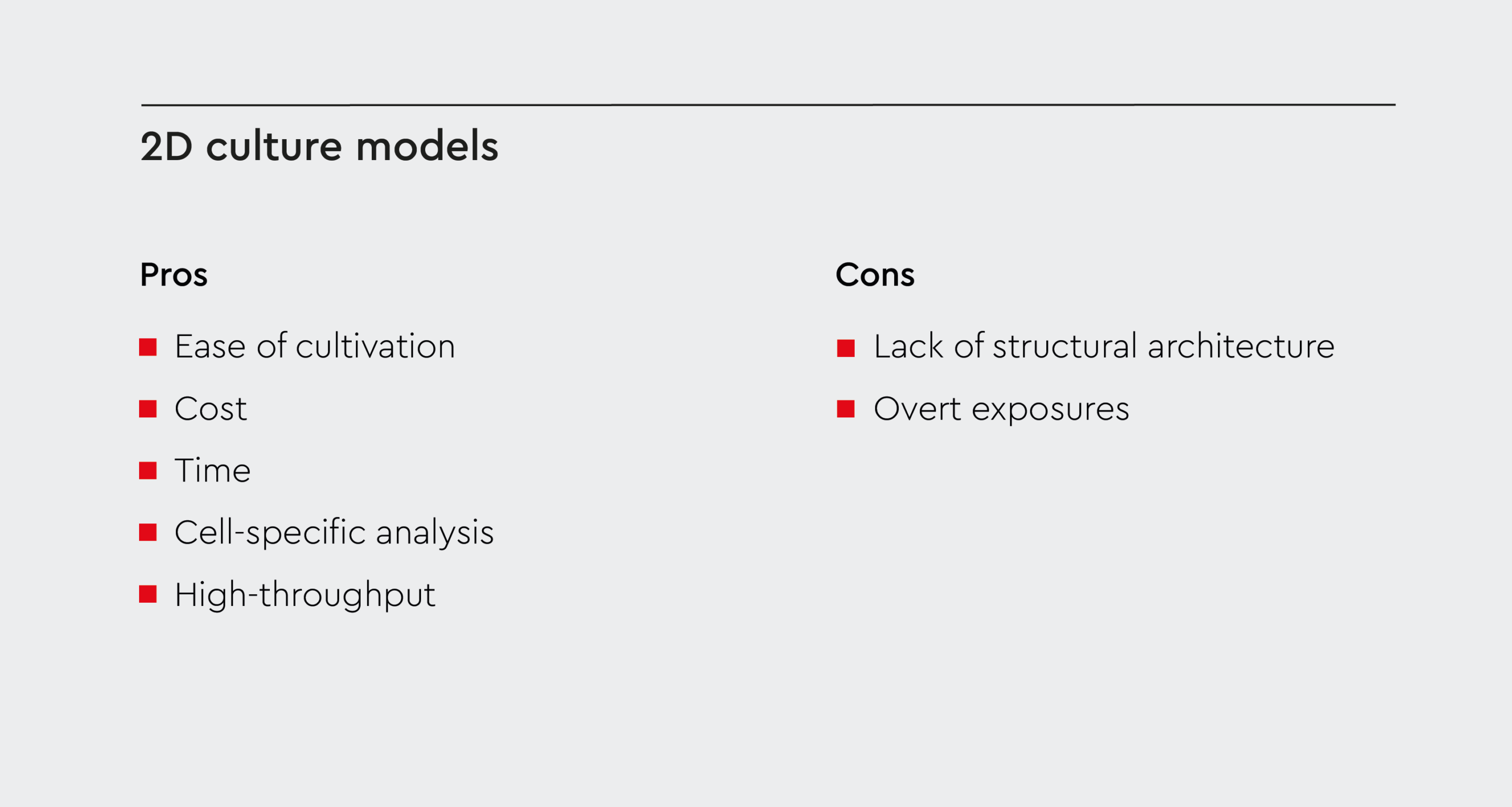
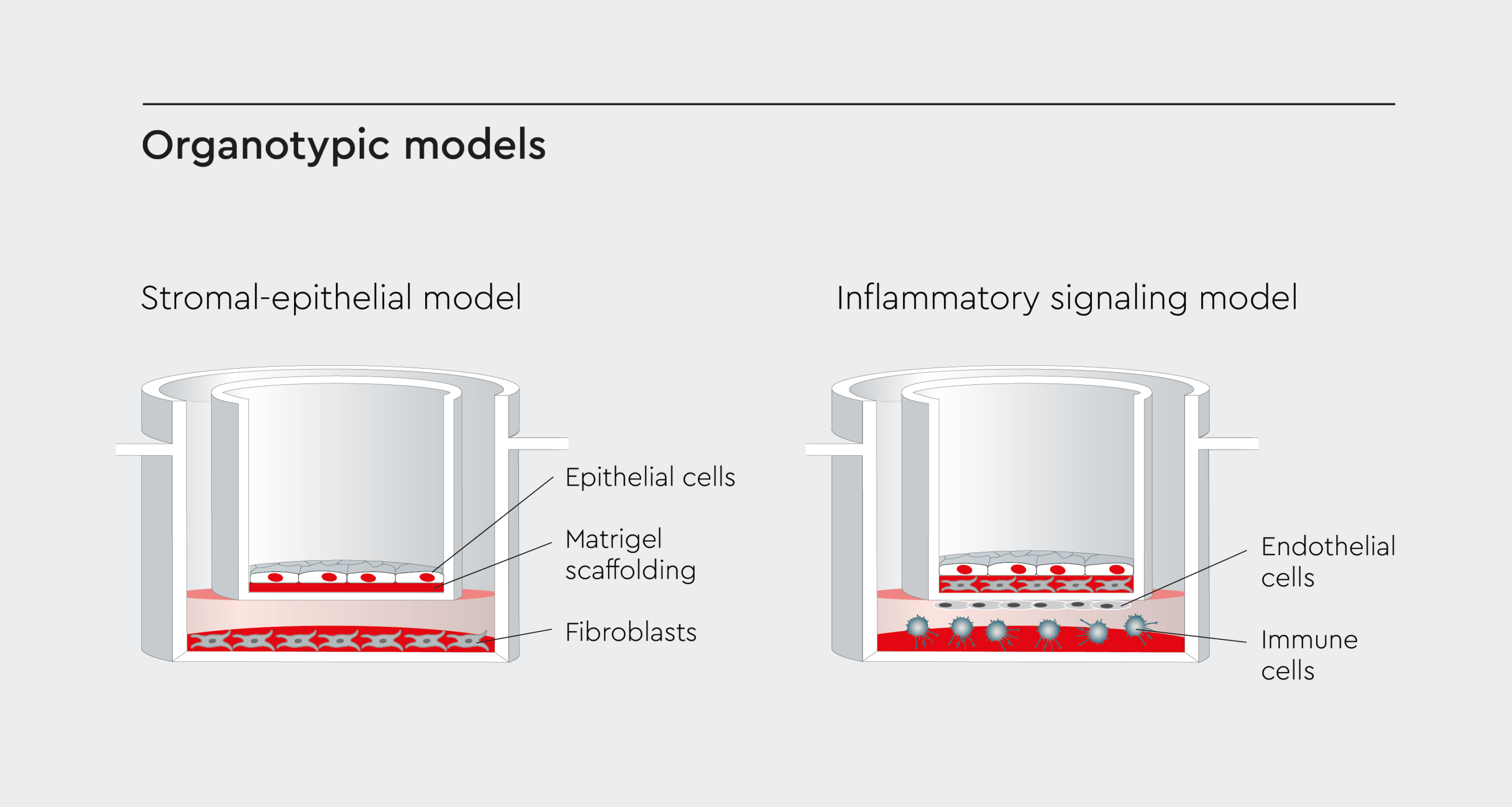
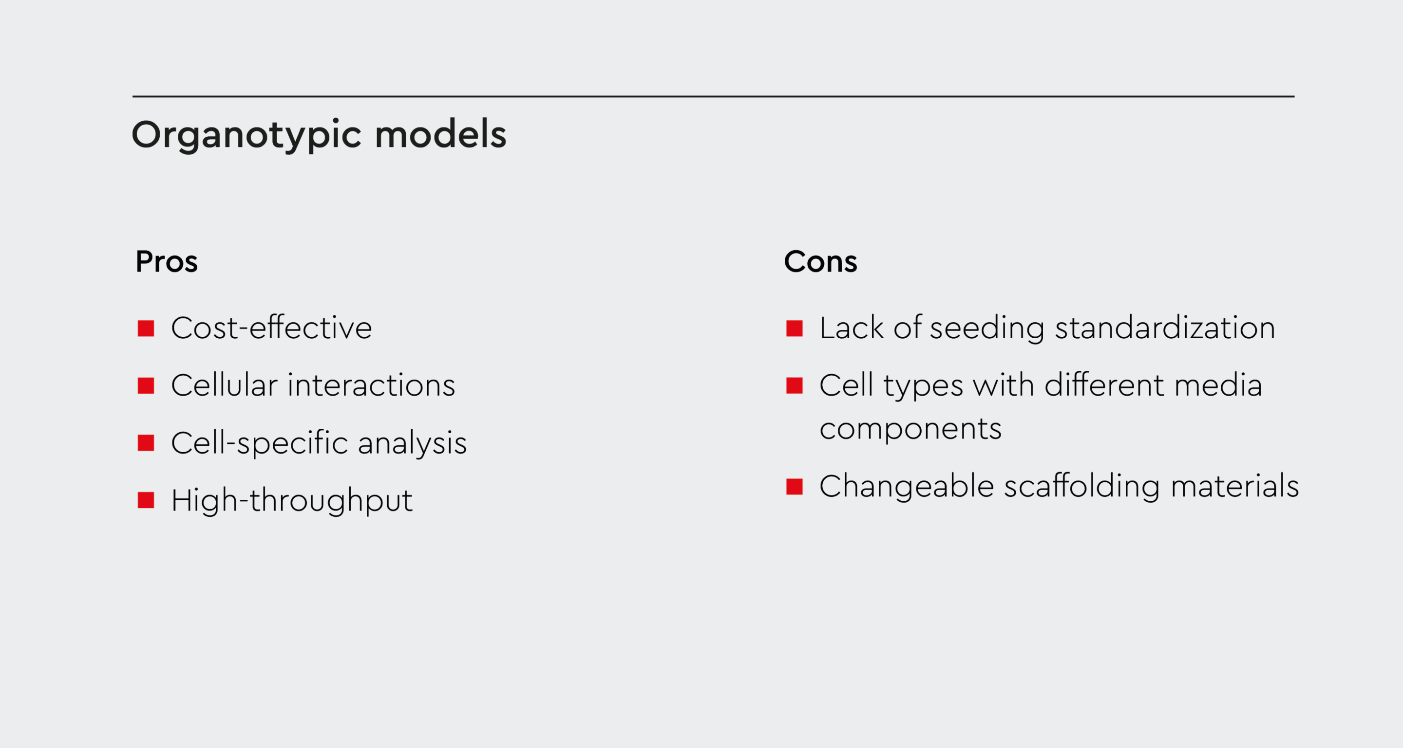
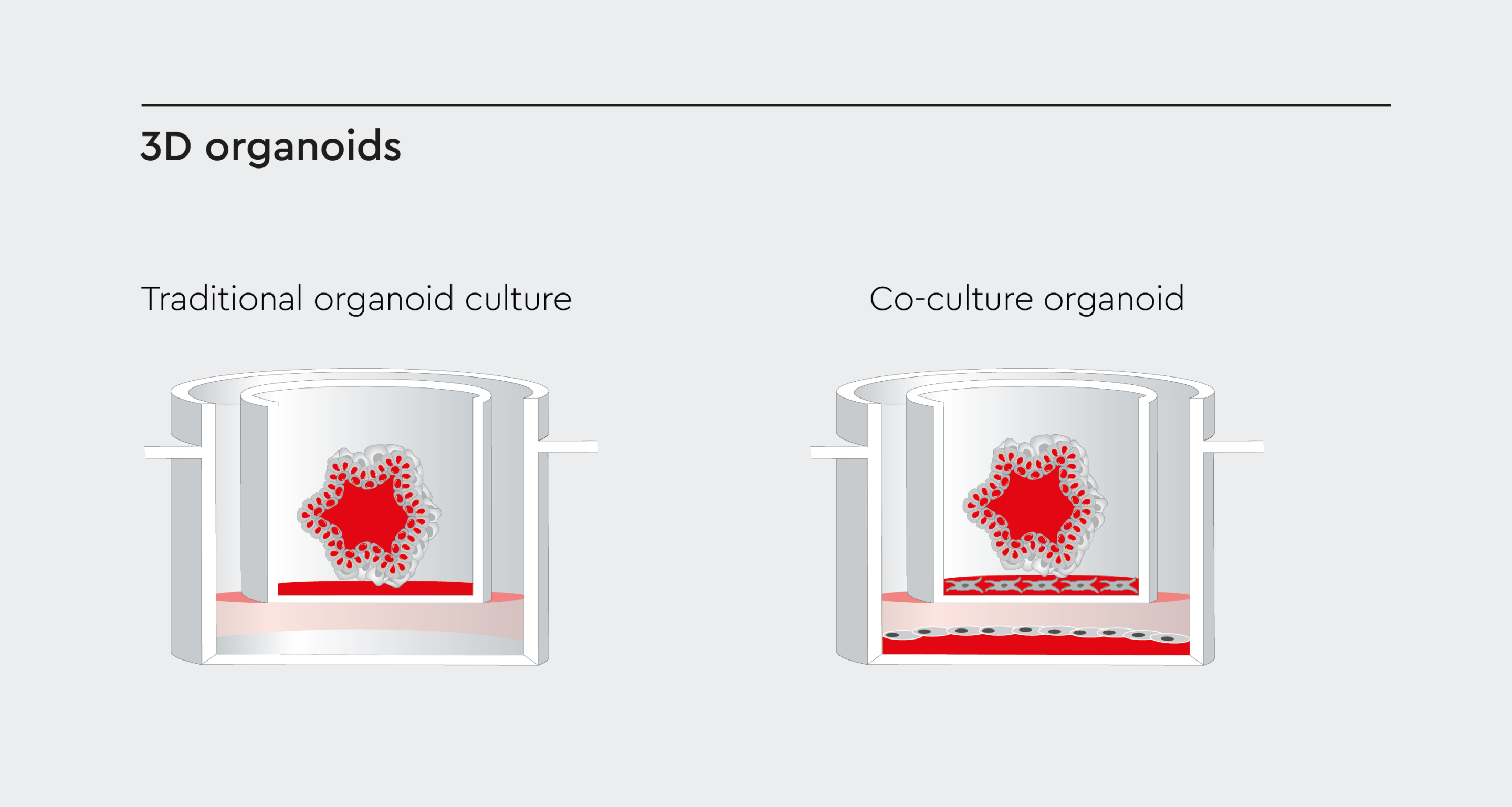
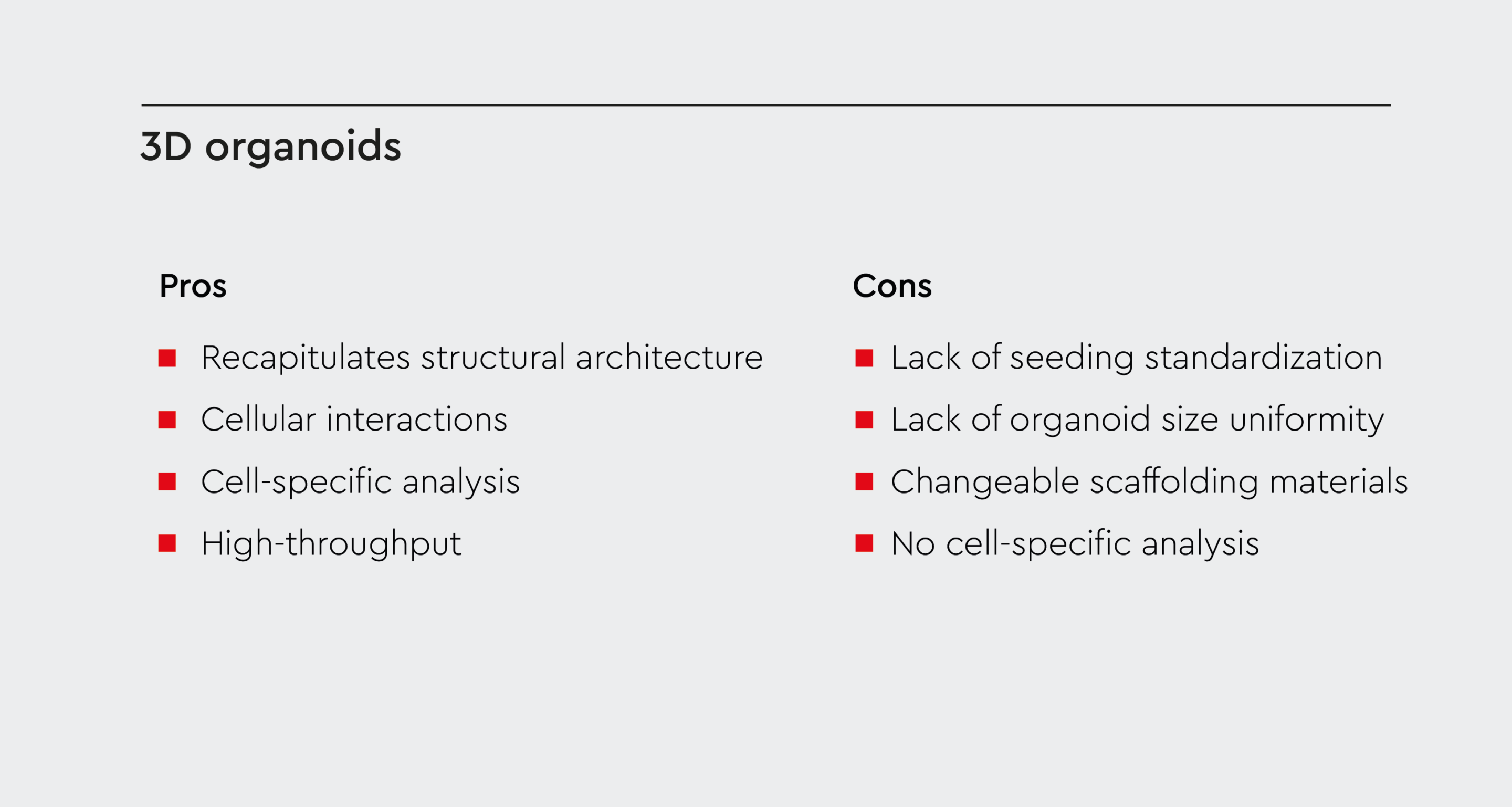
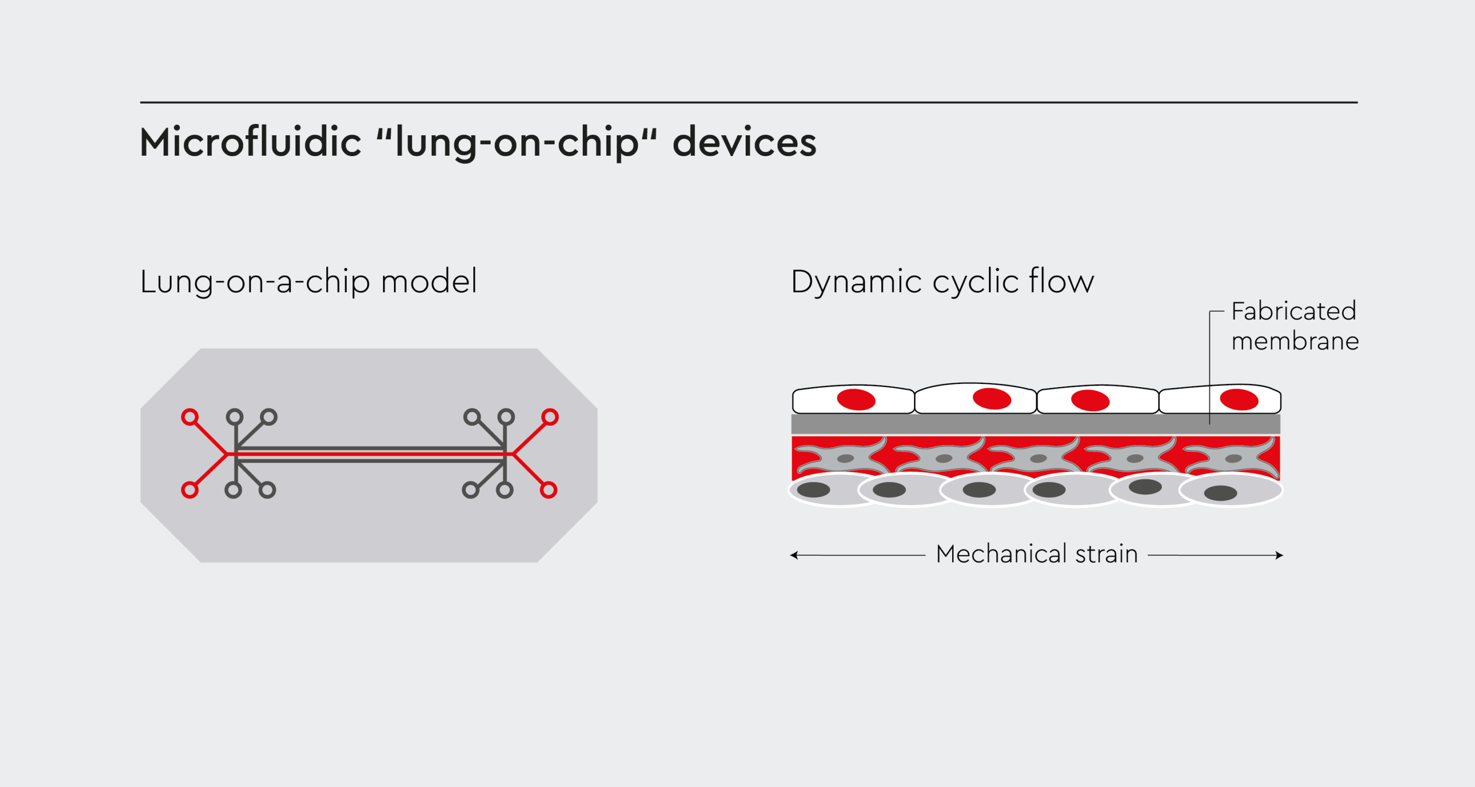
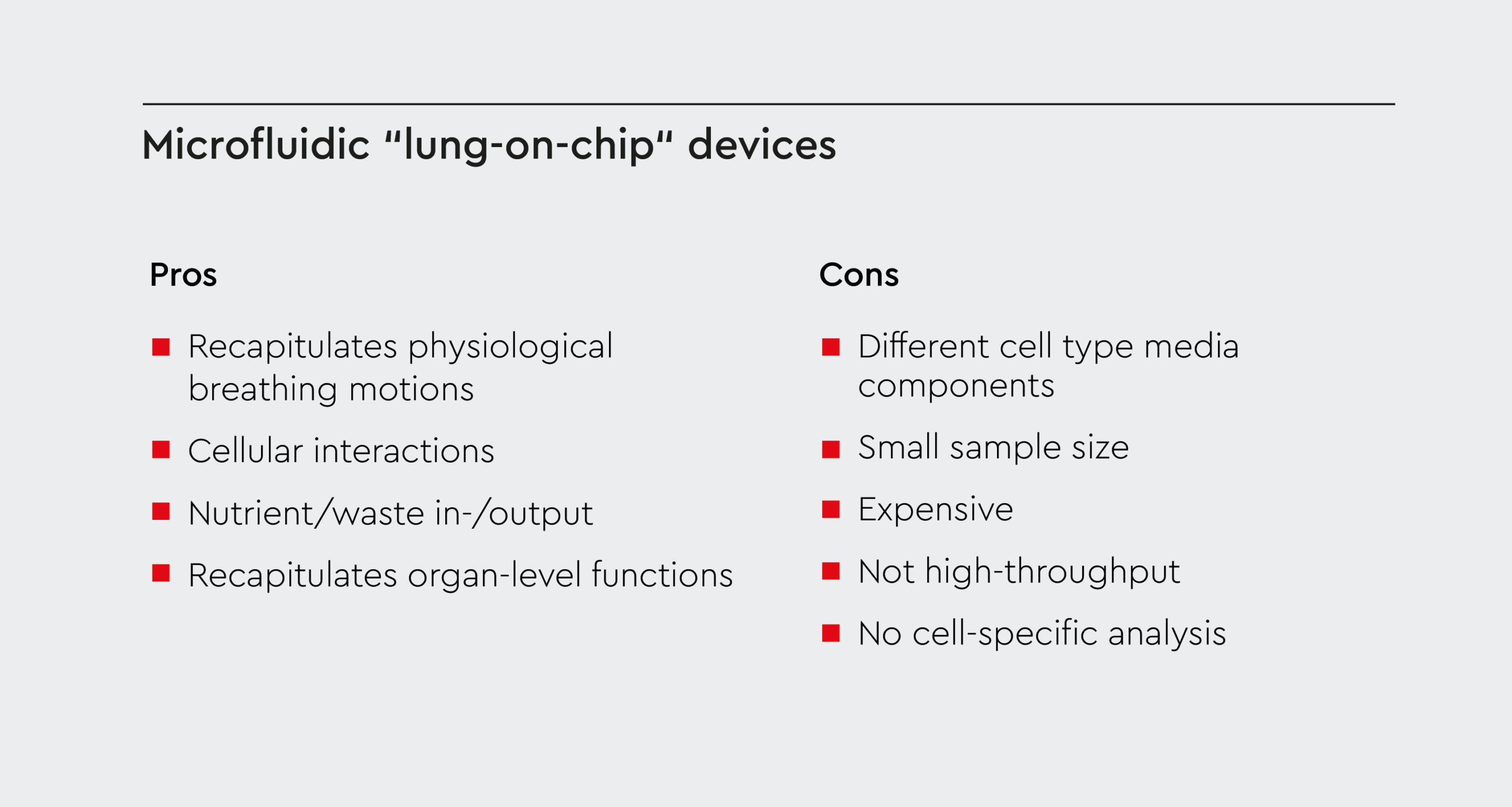
Figure 2: Diagram showing different types of in vitro lung models
Adapted from Faber et al., 2018.
The promise of in vitro lung models
In vitro models hold immense promise for respiratory research and offer several advantages over animal models:- Precise control over experimental conditions: In vitro lung models allow researchers to control variables with precision, providing a level of control that is not feasible in animal models.7
- Reduced reliance on animal models: Utilizing in vitro models decreases the need for animal testing, aligning with ethical considerations and reducing costs.8
- Reproducibility and scalability: These models offer reproducibility for rigorous scientific research and scalability for high-throughput studies.9
- Mimicking physiological conditions: Unlike animal models that poorly mimic human lung biology, in vitro models developed using human cells closely mimic the physiological conditions of the human lung.10
- Versatility: Unlike animal models, they can easily be adjusted based on experimental needs or tailored to mimic specific features of human lung disease.11
- Scalability: Drug screening often requires large amounts of cells, and in vitro lung models allow for high-throughput testing of drugs.12
Importance of in vitro lung models
The importance of in vitro lung models as accurate and reliable models for studying lung biology and diseases transcends disciplinary boundaries. From respiratory infections, such as COVID-19, to non-small cell lung cancer and other lung malignancies, lung diseases pose a heavy burden on patients and healthcare systems (Figure 3). The use of in vitro models to gain a better understanding of these diseases is instrumental in the development of effective and potentially life-saving therapies.- Lung biology studies: These models help understand the fundamental mechanisms of lung function. They can be used to study the process of lung development, the role of different cell types in the lung, and the response of the lung to various stimuli.4
- Respiratory disease research: Researchers use in vitro lung models to gain a deeper understanding of lung diseases like asthma, chronic obstructive pulmonary disease, lung cancer, and COVID-19. They can be used to model disease mechanisms, identify disease biomarkers, and study the effects of potential treatments.4 In vitro lung models are also important for understanding the pathogenic mechanisms of new airborne viruses, such as the SARS-CoV-2 virus, and developing novel prevention strategies and therapies for respiratory infections that have the potential to turn into epidemics or pandemics.13
- Drug discovery and testing: In vitro respiratory models provide a platform for testing the efficacy and safety of novel drugs. They can be used for high-throughput screening of drug candidates or to study drug absorption, distribution, metabolism, and excretion.12
- Environmental exposure studies: In vitro lung models help in studying the effects of environmental pollutants on lung health. They can be used to model the effects of smoking, pathogens, and occupational exposures on the lung.12
- Personalized medicine: These models can be used to create patient-specific lung models to study disease mechanisms and test treatments.14 This can help in the development of personalized treatment strategies.
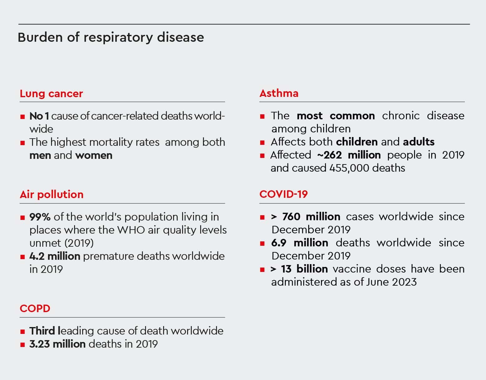
Figure 3: Burden of respiratory disease
Data obtained from the World Health Organization (WHO) website (https://www.who.int/).
Shaping the future of respiratory research
Challenges of in vitro lung model research
Despite the numerous advantages of in vitro models over animal models of respiratory diseases, there are also challenges associated with using in vitro lung models:- Complexity of replicating cellular interactions: Despite advancements, accurately replicating the intricate cellular interactions within the lung remains challenging.15
- Limited representation of systemic effects: In vitro models may not fully capture the systemic effects seen in whole organisms.16
- Standardization across models and laboratories: Achieving standardization across different in vitro lung models remains a challenge, impacting result reproducibility.17
Improving in vitro models for respiratory research
Recent advances in bioengineering, material science, and microfluidics have led to significant improvements in in vitro respiratory models. Advances include innovative culture systems and enhanced media formulations to improve the reproducibility of lung model development.18 Culture at an air interface induces epithelial differentiation to form distinct tracheal, bronchial, or alveolar phenotypes depending on the medium formulation.19 Advanced techniques such as air-liquid interface (ALI) cultures and the use of decellularized lung scaffolds and biomimetic matrixes have enabled the development of more realistic lung models that contain distinct respiratory tract domains.19 Additional advancements include the incorporation of advanced biomaterials to mimic the extracellular matrix of the lung, the development of microfluidic systems to simulate breathing motions, and the integration of advanced imaging techniques for real-time monitoring of cellular responses.20,21 The evolution of in vitro lung models signifies a paradigm shift in respiratory research. Continuous advancements in culture systems, improved media formulations, and sophisticated imaging techniques, combined with an ever-expanding understanding of lung biology, pave the way for more sophisticated and reliable models that bridge the gap between traditional models and the complexity of the human lung. The future of in vitro lung models is promising, with ongoing research focused on improving the physiological relevance of these models and their applicability in personalized medicine.Our innovative solutions for in vitro lung
We provide a comprehensive portfolio of primary cells and specialized cell culture media that can aid in the establishment of in vitro lung models (Figure 1). Our cutting-edge products include:- Human Bronchial Epithelial Cells (HBEpC): These cells are ideal for creating bronchial models. They can be cultured using our Airway Epithelial Cell Growth Medium or Air-Liquid Interface Medium.
- Human Pulmonary Fibroblasts (HPF): HPF cells are crucial for studying the role of fibroblasts in lung diseases. They can be grown using our Fibroblast Growth Medium 2.
- Human Small Airway Epithelial Cells (HSAEpC): These cells are useful for creating models of the small airways and can be cultured using our Small Airway Epithelial Cell Growth Medium.
References
Expand
- Tan CL, Chan Y, Candasamy M, et al. Unravelling the molecular mechanisms underlying chronic respiratory diseases for the development of novel therapeutics via in vitro experimental models. Eur J Pharmacol. 2022;919:174821.
- Nossa R, Costa J, Cacopardo L, Ahluwalia A. Breathing in vitro: Designs and applications of engineered lung models. J Tissue Eng. 2021;12:20417314211008696.
- Rock JR, Hogan BLM. Epithelial progenitor cells in lung development, maintenance, repair, and disease. Annu Rev Cell Dev Biol. 2011;27:493-512.
- Miller AJ, Spence JR. In vitro models to study human lung development, disease and homeostasis. Physiology. 2017;32(3):246-260.
- Petpiroon N, Netkueakul W, Sukrak K, et al. Development of lung tissue models and their applications. Life Sci. Published online 2023:122208.
- Bennet TJ, Randhawa A, Hua J, Cheung KC. Airway-On-A-Chip: designs and applications for lung repair and disease. Cells. 2021;10(7):1602.
- Sakagami M. In vivo, in vitro and ex vivo models to assess pulmonary absorption and disposition of inhaled therapeutics for systemic delivery. Adv Drug Deliv Rev. 2006;58(9-10):1030-1060.
- Cheluvappa R, Scowen P, Eri R. Ethics of animal research in human disease remediation, its institutional teaching; and alternatives to animal experimentation. Pharmacol Res Perspect. 2017;5(4):e00332.
- Baran SW, Brown PC, Baudy AR, et al. Perspectives on the evaluation and adoption of complex in vitro models in drug development: Workshop with the FDA and the pharmaceutical industry (IQ MPS Affiliate). ALTEX-Alternatives to animal experimentation. 2022;39(2):297-314.
- Gordon S, Daneshian M, Bouwstra J, et al. Non-animal models of epithelial barriers (skin, intestine and lung) in research, industrial applications and regulatory toxicology. ALTEX. 2015;32(4):327-378.
- Bonniaud P, Fabre A, Frossard N, et al. Optimising experimental research in respiratory diseases: an ERS statement. European Respiratory Journal. 2018;51(5).
- Astashkina A, Mann B, Grainger DW. A critical evaluation of in vitro cell culture models for high-throughput drug screening and toxicity. Pharmacol Ther. 2012;134(1):82-106.
- Heinen N, Kloehn M, Steinmann E, Pfaender S. In vitro lung models and their application to study SARS-CoV-2 pathogenesis and disease. Viruses. 2021;13(5):792.
- Kim M, Mun H, Sung CO, et al. Patient-derived lung cancer organoids as in vitro cancer models for therapeutic screening. Nat Commun. 2019;10(1):3991.
- Doryab A, Tas S, Taskin MB, et al. Evolution of bioengineered lung models: recent advances and challenges in tissue mimicry for studying the role of mechanical forces in cell biology. Adv Funct Mater. 2019;29(39):1903114.
- BéruBé K, Prytherch Z, Job C, Hughes T. Human primary bronchial lung cell constructs: the new respiratory models. Toxicology. 2010;278(3):311-318.
- Faber SC, McCullough SD. Through the looking glass: in vitro models for inhalation toxicology and interindividual variability in the airway. Appl In Vitro Toxicol. 2018;4(2):115-128.
- Badr-Eldin SM, Aldawsari HM, Kotta S, Deb PK, Venugopala KN. Three-Dimensional in vitro cell culture models for efficient drug discovery: Progress so far and future prospects. Pharmaceuticals. 2022;15(8):926.
- Kumar P, Vrana NE, Ghaemmaghami AM. Prospects and challenges in engineering functional respiratory epithelium for in vitro and in vivo applications. Microphysiol Syst. 2017;1.
- Shao Y, Fu J. Integrated micro/nanoengineered functional biomaterials for cell mechanics and mechanobiology: a materials perspective. Advanced Materials. 2014;26(10):1494-1533.
- Ding S, Zhang H, Wang X. Microfluidic-chip-integrated biosensors for lung disease models. Biosensors (Basel). 2021;11(11):456.
Contact our experts As a leader in developing cell culture solutions, we recognize that the future of respiratory research relies on innovative in vitro cell culture models of the human lung. Our commitment to providing high-quality cells and media ensures that researchers have the tools they need to advance our understanding of respiratory diseases.
If you're a researcher or clinical scientist working in respiratory research, contact our specialists today to discover how our human primary cells and specialized cell culture media can help you advance your respiratory research.