Cell culture experiments form the backbone of biomedical research and drug development. However, phenol red, a common component of cell culture media, can influence the results of in vitro assays. In this article, we explore the limitations of using phenol red in cell culture and introduce innovative phenol red-free (prf) alternatives that promise enhanced experimental accuracy and reproducibility. Read on to learn more about how switching to prf media can improve the reliability of your research.
What is the function of phenol red in cell culture?
Phenolsulfonphthalein, commonly referred to as phenol red, has been a standard component of cell culture media for several decades. This organic compound serves as a pH indicator, changing color in response to pH variations. Its ability to change color in response to pH variations is due to its molecular structure, which undergoes conformational changes in different pH environments.1 At physiological pH (between pH 6.8 and 8.2), phenolsulfonphthalein exhibits its characteristic red color (Figure 1). When the medium becomes acidic (pH 6.4 or lower), it turns yellow, signaling potential bacterial contamination or excessive cellular metabolism. Conversely, a shift to purple (pH 8.0 or higher) may indicate alkaline conditions or contamination with alkaline substances.1–3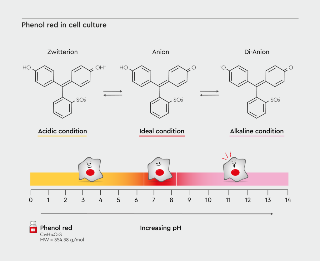
Figure 1: pH-dependent color changes of phenol red in cell culture.
Visual representation showing the color spectrum along the spectrum of pH levels, from yellow (acidic) to pink (alkaline). Adapted from Weiskirchen et al., 2023.1
Limitations of phenol red-containing culture media
Although phenol red in cell culture allows for quick and easy pH monitoring, its presence can influence the results of cell culture experiments in several ways:- Estrogenic activity: It exhibits structural similarity to certain steroid hormones and possesses weak estrogenic properties that can affect hormone-sensitive cells. Therefore, the presence of phenol red in cell culture could skew results in endocrine research and studies involving xenoestrogens or breast cancer cells.5,6
- Fluorescence interference: It can interfere with fluorescence-based assays by contributing to background fluorescence or by absorbing light at specific wavelengths.4 This spectral interference can affect the results of flow cytometry, fluorescence microscopy, and other fluorescence-based assays.
- Potential cytotoxicity: Phenol red is generally considered non-toxic at the concentrations used in cell culture media. However, some studies have suggested that it may have cytotoxic effects on certain cell types, potentially impacting cell growth and experimental results.7 The sensitivity to phenol red appears to vary among different cell types, with some cell types (e.g., breast cancer cells) showing more pronounced effects than others, adding another layer of complexity to experimental design and interpretation.8
- Cell-specific effects: Some studies have shown that the presence of phenol red in cell culture can influence cell proliferation rates, protein expression patterns, and cellular responses to various stimuli.4,9,10 These effects vary among cell types and experimental conditions, adding an unwanted variable to research protocols.
- Assay interference: Phenolsulfonphthalein can affect colorimetric and spectrophotometric measurements, potentially compromising experimental accuracy.2,11 This interference can lead to false positives or negatives, potentially affecting drug development pipelines and research findings.
Moving beyond phenol red in cell culture research
The transition to a phenol red-free environment necessitates alternative methods for monitoring culture conditions. Modern laboratories employ various strategies, including:- Separate pH meters with specialized probes for periodic measurements12
- Other pH indicators with different spectral properties, such as bromothymol blue and m-Cresol purple13
- Advanced monitoring systems integrated with incubators14
The clear solution: Using media without phenol red
The most straightforward and increasingly popular solution is the use of cell culture media without phenolsulfonphthalein. These formulations maintain the necessary nutrients for cell growth without the potential interference. Advantages of phenol red-free media include:- Elimination of potential hormone-like effects, allowing for more accurate studies of hormone-responsive pathways and improved reliability in endocrine disruption research.4,5
- Improved assay accuracy and reproducibility, providing more consistent results across different experimental setups and enhanced sensitivity for detecting subtle cellular responses.5,15
- Greater flexibility in experimental design, providing improved compatibility with a wide range of assays and greater control over the chemical composition of the culture environment.4,10
- Reduced interference in optical and fluorescence-based techniques, allowing for clearer imaging of cellular structures and processes and improved performance in high-throughput screening applications.4
- Regulatory compliance in biopharmaceutical manufacturing, reducing the potential for unexpected interactions between media components and the final product.5,16
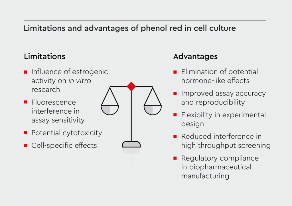
Figure 2: Limitations and advantages of phenol red in cell culture.
The scheme illustrates the key limitations of phenol red-containing media (left) and the benefits of switching to phenol red-free formulations (right).
Applications of phenol red-free media
Media without phenolsulfonphthalein are ideal for use in various research areas and experimental setups:- Hormone-dependent studies, including breast cancer research and endocrine disruption investigations.5
- Fluorescence-based experiments, including high-resolution fluorescence microscopy, flow cytometry, high-content screening, and live cell imaging.4
- Response tracking studies, including drug screening, toxicology investigations, and cellular metabolism studies.5
- Stem cell research, including differentiation studies and regenerative medicine applications.17
- Manufacturing of biopharmaceuticals under strict regulatory guidelines, including production of recombinant proteins, manufacturing of cell-based therapies, and development of vaccines.17
Cell culture solutions without phenol red
As cell culture experts, we recognize the growing demand for cell culture media without phenol red. We offer an extensive range of ready-to-use prf formulations designed for diverse research needs:- Cryo-SFM Plus phenol red-free provides superior cryopreservation performance for sensitive cell types. The formulation ensures high post-thaw viability and maintains cellular characteristics during long-term storage.
- Mesenchymal Stem Cell Growth Medium XF supports robust expansion of mesenchymal stem cells while maintaining their multipotency. This specialized formulation enables maintenance of the self-renewal ability and differentiation potential of stem cells.
- Hematopoietic Progenitor Cell Expansion Medium XF facilitates optimal growth and maintenance of hematopoietic stem and progenitor cells, which are crucial for blood cell development research.
- Mesenchymal Stem Cell Growth Medium 2 ensures the robust expansion of mesenchymal stem cells from various sources and maintains their marker expression and differentiation capacity.
- Pericyte Growth Medium 2 supports optimal growth and maintenance of pericytes. This formulation preserves pericyte-specific markers and functionality.
- 3D Tumorsphere Medium XF supports the formation and growth of cancer stem cell-enriched spheroids and enables studies of tumor initiation, progression, and drug resistance. This prf version of this medium is particularly valuable for breast cancer research, where the absence of phenol red’s estrogenic properties ensures more reliable results in studying cancer stem cells and therapeutic responses.
References
Expand
-
- Weiskirchen S, Schröder SK, Buhl EM, Weiskirchen R. A beginner’s guide to cell culture: practical advice for preventing needless problems. Cells. 2023;12(5):682. doi:10.3390/cells12050682
- Raffay R, Husin N, Omar AF. Spectrophotometry and colorimetry profiling of pure phenol red and cell culture medium on pH variation. Color Technol. 2022;138(6):640-659. doi:10.1111/cote.12626
- Louis KC, Hamid SHFWA, Raffay R, Omar AF. Specialized colorimeter for phenol red pH measurement. J Phys Conf Ser. 2022;2411(1):012020. doi:10.1088/1742-6596/2411/1/012020
- Sundarakrishnan A, Herrero Acero E, Coburn J, Chwalek K, Partlow B, Kaplan DL. Phenol red-silk tyrosine cross-linked hydrogels. Acta Biomater. 2016;42:102-113. doi:10.1016/j.actbio.2016.06.020
- Berthois Y, Katzenellenbogen JA, Katzenellenbogen BS. Phenol red in tissue culture media is a weak estrogen: implications concerning the study of estrogen-responsive cells in culture. Proc Natl Acad Sci U S A. 1986;83(8):2496-2500.
- Liu X, Chen B, Chen L, et al. U-shape suppressive effect of phenol red on the epileptiform burst activity via activation of estrogen receptors in primary hippocampal culture. PLOS ONE. 2013;8(4):e60189. doi:10.1371/journal.pone.0060189
- Zhu Y, Zhang X, Zhu J, et al. Cytotoxicity of phenol red in toxicity assays for carbon nanoparticles. Int J Mol Sci. 2012;13(10):12336-12348. doi:10.3390/ijms131012336
- Grady LH, Nonneman DJ, Rottinghaus GE, Welshons WV. pH-dependent cytotoxicity of contaminants of phenol red for MCF-7 breast cancer cells. Endocrinology. 1991;129(6):3321-3330. doi:10.1210/endo-129-6-3321
- Lephart ED. 4′,7-isoflavandiol (Equol) enhances human dermal fibroblast renewal and has effects similar to 17β-estradiol in stimulating collagen and elastin expression. Cell cycle and RT-PCR analysis without phenol red. Cosmetics. 2021;8(1):5. doi:10.3390/cosmetics8010005
- Węsierska-Gądek J, Schreiner T, Maurer M, Waringer A, Ranftler C. Phenol red in the culture medium strongly affects the susceptibility of human MCF-7 cells to roscovitine. Cell Mol Biol Lett. 2007;12(2):280-293. doi:10.2478/s11658-007-0002-5
- Foreman RE, Lucey R, Leaney AR, Lee MY, Naseer H, Wilson A. Optimized LC-MS/MS methods for quantifying antibody–drug conjugate payloads in cell culture media containing phenol red. Bioanalysis. 2024;16(12):575-585. doi:10.1080/17576180.2024.2349422
- Dean C, Gunasegaran K, Gabriel J, Iacobelli D, Sorby K. pHantastic embryos and where to find them: can a real-time pH monitoring system represent the pH environment of culture media? Fertil Reprod. 2022;04(03n04):181-181. doi:10.1142/S2661318222740929
- Nguyen DK, Nguyen HQ, Dang HTT, Nguyen VQ, Nguyen L. A low-cost system for monitoring pH, dissolved oxygen and algal density in continuous culture of microalgae. HardwareX. 2022;12. doi:10.1016/j.ohx.2022.e00353
- Casimero C, McConville A, Fearon JJ, et al. Sensor systems for bacterial reactors: a new flavin-phenol composite film for the in situ voltammetric measurement of pH. Anal Chim Acta. 2018;1027:1-8. doi:10.1016/j.aca.2018.04.053
- Guzik K, Reiss K, Korohoda W. Survival and phenol red metabolism in chick embryo hepatocytes cultured in the serum-free medium 199 supplemented with dextran T-500 and antioxidants. Folia Histochem Cytobiol. 1989;27(3):125-130.
- Glover JF, Irwin JT, Darbre PD. Interaction of phenol red with estrogenic and antiestrogenic action on growth of human breast cancer cells ZR-75-1 and T-47-D. Cancer Res. 1988;48(13):3693-3697.
- Morgan A, Babu D, Reiz B, Whittal R, Suh LYK, Siraki AG. Caution for the routine use of phenol red – It is more than just a pH indicator. Chem Biol Interact. 2019;310:108739. doi:10.1016/j.cbi.2019.108739
Contact our experts
Ready to enhance your cell culture research? Contact our specialists to discover how our phenol red-free solutions can elevate your research quality and to help you select the optimal formulation for your specific needs.
Contact us