
Adipocytes are mature fat cells that help regulate metabolic homeostasis. They store and release energy, secrete signalling molecules, and contribute to whole-body processes such as lipid metabolism, glucose regulation, inflammation, and thermogenesis.
As the main cellular component of adipose tissue, adipocytes do far more than passively store lipids. They respond to nutritional, hormonal, and inflammatory cues, helping the body adapt to changing energy demands.
When adipocyte function is disrupted, adipose tissue can become a driver of metabolic imbalance. Impaired lipid storage, chronic low-grade inflammation, and altered adipokine secretion are closely linked to obesity, insulin resistance, type 2 diabetes, and cardiovascular disease.
To learn more about the relationship between obesity and type 2 diabetes, visit our diabetes research overview.
For researchers, this makes adipocytes highly relevant models for understanding how metabolic homeostasis is maintained, how it breaks down in disease, and how potential therapeutic strategies may be explored in vitro.
In this article, we look at the different types of adipocytes, their role in energy storage and endocrine signalling, and how adipocyte dysfunction contributes to metabolic disease.
What are adipocytes and what does adipose tissue do?
Adipose tissue is a highly dynamic organ involved in energy storage, endocrine signalling, immune regulation, and metabolic balance. Its main cellular component is the adipocyte, a mature fat cell specialized in storing lipids and responding to changes in energy demand.
Acting as an energy bank, storing and releasing energy is just one role of adipose tissue. Adipocytes also produce and secrete hormones and signalling molecules that influence appetite, inflammation, insulin sensitivity, cardiovascular activity, and tissue regeneration.
Adipocytes are not the only components of adipose tissue. The tissue also contains preadipocytes, macrophages, fibroblasts, endothelial cells, connective tissue, and stem cells. Together, these cells help maintain adipose tissue structure, adipocyte integrity, and hormonal balance.
To support studies in this field, we offer adipose tissue-derived cells and media for adipose tissue research.
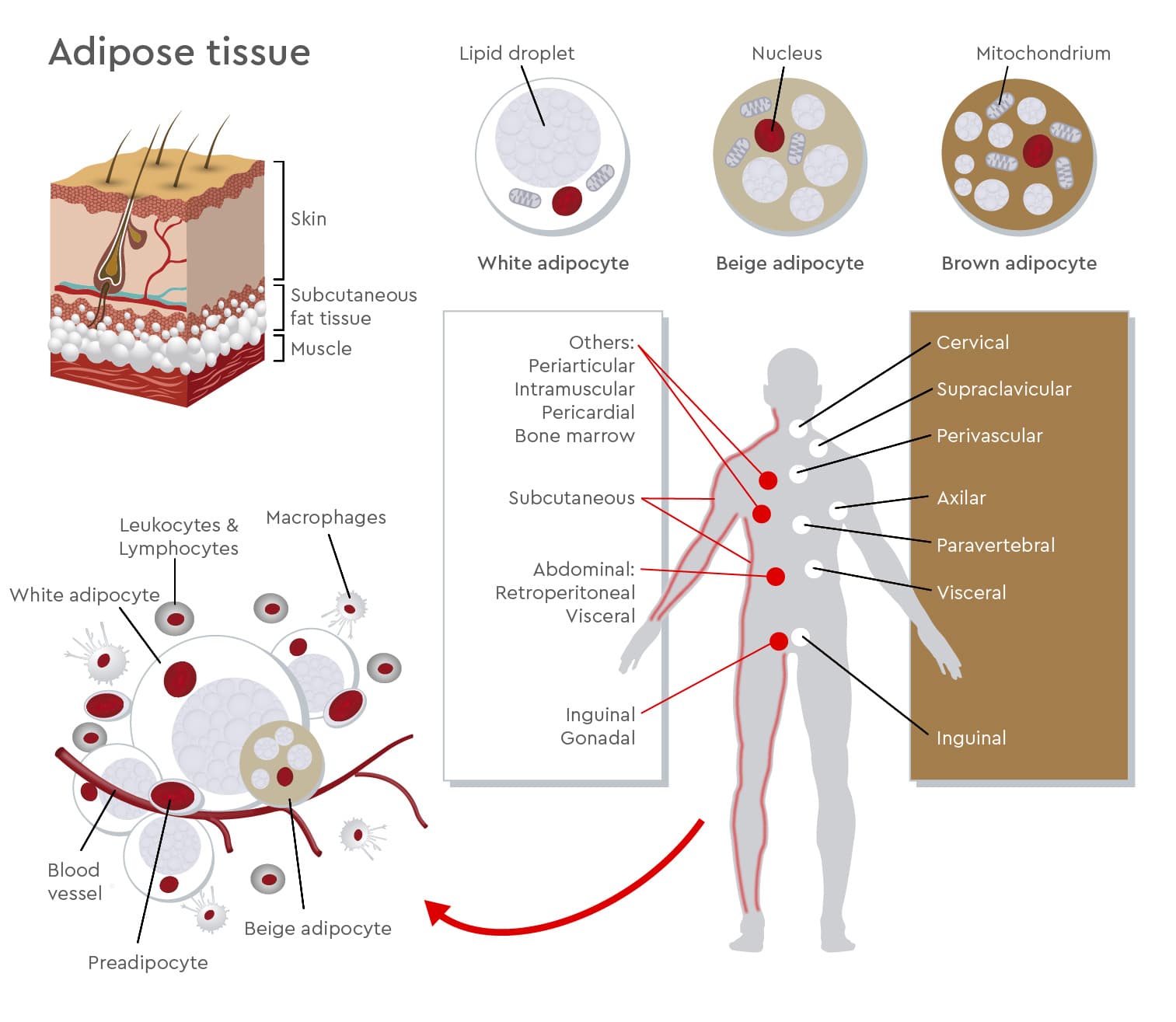
Figure 1: Adipose tissue structure and adipocyte diversity.
Adipose tissue contains mature adipocytes together with preadipocytes, immune cells, macrophages, blood vessels, and other stromal cell populations. White, beige, and brown adipocytes differ in lipid droplet structure, mitochondrial content, and metabolic function. These adipocyte types are found across different anatomical depots and contribute to energy storage, endocrine signalling, thermogenesis, and metabolic regulation.
What are the main types of adipocytes?
Humans have different adipocyte types with distinct but partly overlapping functions. White adipocytes primarily support energy storage and endocrine signalling, brown adipocytes are specialized for heat production, and beige or brite adipocytes can acquire thermogenic, brown-like features in response to specific stimuli.
White adipocytes are large cells that usually contain one dominant lipid droplet and fewer mitochondria than thermogenic adipocytes. They store excess energy as triglycerides and release fatty acids when energy is needed. White adipose tissue also acts as an endocrine organ, secreting adipokines, cytokines, and other signalling molecules involved in appetite regulation, inflammation, insulin sensitivity, cardiovascular activity, and tissue remodelling.1
Brown adipocytes are smaller, mitochondria-rich cells with several lipid droplets. They generate heat through thermogenesis and contribute to energy expenditure. Beyond heat production, brown adipocytes also secrete signalling molecules that may influence local and systemic metabolism.2
Beige, or brite, adipocytes are inducible thermogenic adipocytes found within white adipose tissue depots. At baseline, they can resemble white adipocytes, but after stimulation, such as cold exposure or adrenergic signalling, they may acquire brown-like features, including higher mitochondrial activity and thermogenic capacity.3 This reflects the plasticity of adipose tissue rather than a fixed, one-directional cell identity.
| Feature | White adipocytes | Brown adipocytes | Beige/brite adipocytes |
|---|---|---|---|
| Main function | Energy storage and endocrine signalling | Heat production | Inducible thermogenesis |
| Lipid droplets | One large lipid droplet | Several small lipid droplets | White-like at rest; brown-like after activation |
| Mitochondria | Fewer mitochondria | Many mitochondria | Increase with activation |
| Research relevance | Obesity, inflammation, insulin resistance | Energy expenditure and thermogenesis | Browning, metabolic adaption, therapeutic research |
Table 1. White, brown, and beige adipocytes differ in lipid droplet structure, mitochondrial content, and metabolic function. Together, they contribute to energy storage, endocrine regulation, and thermogenesis.
How do adipocytes support metabolic homeostasis?
Adipocytes support metabolic homeostasis by coordinating energy storage, energy release, endocrine signalling, and inflammatory responses. Through these functions, adipose tissue helps the body adapt to changes in nutrient availability and energy demand.
White adipocytes store excess energy as triglycerides and release fatty acids when energy is needed. At the same time, adipocytes secrete adipokines and cytokines that influence appetite, insulin sensitivity, inflammation, cardiovascular function, and tissue remodelling.
Mitochondrial function also contributes to adipocyte biology. It supports adipocyte differentiation and lipid metabolism and is especially important for thermogenesis in brown and beige adipocytes.
How does adipose tissue dysfunction contribute to obesity and insulin resistance?
Adipose tissue dysfunction can contribute to insulin resistance when white adipose tissue can no longer safely store excess energy. Lipids may then accumulate in organs such as the liver, muscle, and heart, promoting local inflammation and impaired insulin signalling.
“Adipose tissue contains many molecules that are involved in processes necessary for maintaining metabolic balance. This is why it plays a crucial role in the onset of metabolic diseases,” explains Melissa Olekson, scientific support specialist at PromoCell.
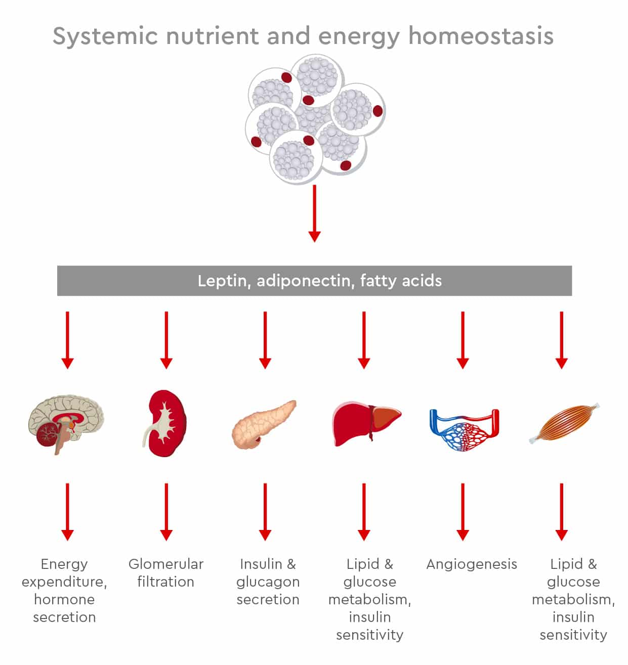
Figure 2: Adipose tissue contributes to systemic nutrient and energy homeostasis by regulating lipid storage, lipid release, endocrine signalling, and inflammatory responses. When these processes become disrupted, adipose tissue dysfunction can affect metabolic organs such as the liver, muscle, heart, and pancreas.
Obesity is a global health challenge and is linked to high-mortality diseases such as type 2 diabetes mellitus and cardiovascular disease. Studies have suggested that 18% of men and 21% of women globally would be classified as obese by 2025, with more than 300 million people suffering from obesity-associated type 2 diabetes.4
Obesity develops when energy intake exceeds energy expenditure. However, it is also shaped by the interaction of many factors, including genetics, epigenetics, environment, and lifestyle.5 This complexity helps explain why researchers still face challenges in understanding the underlying disease mechanisms.
In healthy weight gain, white adipose tissue expands by increasing the size of mature adipocytes and by recruiting and differentiating precursor cells, including mesenchymal stem cells. In unhealthy obesity, white adipose tissue becomes dysfunctional and cannot expand properly to store excess energy.
As a result, fat may be deposited in non-adipose tissues, including liver, muscle, heart, and other visceral organs. This process, known as lipotoxicity, can induce insulin resistance and increase the risk of type 2 diabetes and cardiovascular disease.6
When adipose tissue expands rapidly, it can also trigger cell death, hypoxia, and mechanical stress. These signals promote macrophage infiltration and inflammatory responses. In adipose tissue from obese patients, researchers have found that up to 40% of cells can be macrophages.7
Chronic low-grade inflammation impairs adipose tissue function. It can hinder adipogenesis, reduce insulin sensitivity, and activate immune responses in organs involved in energy homeostasis. This creates an important link between obesity, inflammation, and insulin resistance.
From adipose tissue expansion to insulin resistance
The relationship between obesity and insulin resistance develops through several connected changes in adipose tissue function. The simplified sequence below shows how impaired adipose tissue expansion can contribute to inflammation, reduced insulin sensitivity, and increased cardiometabolic risk.
- Energy intake exceeds energy expenditure
- White adipose tissue expands
- Adipose tissue storage capacity becomes impaired
- Lipids accumulate in non-adipose tissues
- Local inflammation and macrophage infiltration increase
- Adipogenesis and insulin sensitivity are reduced
- The risk of type 2 diabetes and cardiovascular disease increases
Increasing evidence suggests that mitochondria influence the onset and progression of obesity and related pathologies. Damage to the mitochondrial respiratory chain can compromise adipocyte differentiation.8
Based on this knowledge, researchers continue to investigate the molecular mechanisms responsible for adipose tissue dysfunction. A deeper understanding of these mechanisms may support the development of more targeted therapeutic strategies for obesity-related metabolic disease.
For in vitro studies of these mechanisms, human preadipocytes for obesity and diabetes research can support research into adipogenesis, inflammation, and insulin resistance.
Why are brown and beige adipocytes relevant therapeutic targets?
Brown and beige adipocytes are relevant therapeutic targets because they can increase energy expenditure and support glucose and lipid metabolism. Their thermogenic activity makes them especially interesting for research into obesity, insulin resistance, and metabolic disease.
Alongside strategies aimed at improving adipose tissue health, brown adipose tissue and beige adipocytes show promise as therapeutic targets for obesity. Brown adipose tissue is involved in both energy homeostasis and glucose homeostasis.
Beige adipocytes reside among white adipocytes and can be activated in response to external stimuli such as cold temperatures, exercise, and nutrition. During this browning process, beige adipocytes acquire characteristics of brown adipose tissue and consume energy through heat production.
These stimuli may also promote brown-like features in white adipose tissue, depending on depot, cellular context, and signalling environment. Hormones and signalling factors, including prostaglandins, natriuretic peptides, bone morphogenetic protein (BMP), and vascular endothelial growth factor (VEGF), can regulate brown and beige adipocytes. These factors may increase energy expenditure and improve glucose homeostasis and insulin sensitivity.
Emerging data support the creation of a “metabolic sink” for glucose and triglycerides by promoting the development of beige adipocytes.3 Another therapeutic approach could be to block regulators that impair brown and beige adipocyte function in obese patients, such as transforming growth factor beta (TGF-β).
In some studies, TGF-β neutralizing antibodies protected animals from obesity and insulin resistance.9
How can preadipocytes support metabolic disease research in vitro?
Preadipocytes are useful in vitro models for studying adipocyte differentiation, adipose tissue function, and disease-related changes in human cells. They can help researchers investigate mechanisms involved in obesity, insulin resistance, inflammation, and diabetes.
As adipocyte precursor cells, preadipocytes allow researchers to follow the differentiation process from an undifferentiated cell state towards mature adipocytes. This makes them valuable for studying adipogenesis, lipid accumulation, adipokine secretion, and changes in gene or marker expression.
Melissa Olekson, who supports researchers working with in vitro adipose-cell models, highlights their relevance for metabolic disease research: “Preadipocytes offer a very useful cell model. They not only provide insights in key human signaling pathways, but also offer a platform to test possible treatments in vitro.”
Researchers can also use preadipocytes to compare donor- or disease-associated differences. For example, preadipocytes from donors with diabetes can be compared with cells from healthy donors to investigate differences in intracellular signalling, cytokine release, adipokine profiles, and differentiation capacity.Olekson also points to their use in experimental workflows: “The techniques used in these studies include modification of gene expression and analysis of cell markers. Preadipocytes can also be used as a cell model for diabetes studies or for observing adipogenic differentiation of mesenchymal stem cells.”

Melissa Olekson is a scientific support specialist who helps researchers establish in vitro adipose-cell models for studying molecular processes in metabolic disease.
In one study, Kongsuphol and colleagues co-cultured adipose tissue with immune cells in a microfluidic-based in vitro model.10 Because this approach allows researchers to measure cytokines and generate data on inflammatory reactions and insulin sensitivity, it may support diabetic drug screening.
Together, human preadipocyte and adipocyte models can support research into metabolic dysfunction, adipogenesis, inflammatory signalling, and potential therapeutic approaches. When combined with suitable growth and differentiation media, they provide relevant in vitro systems for obesity- and diabetes-related research.
For more guidance on model selection, read our article on choosing the right human preadipocyte cell model for reliable metabolic assays.
Key terms in adipocyte research
| Term | Meaning |
|---|---|
| Adipocytes | Mature fat cells involved in energy storage, endocrine signalling, and metabolic regulation. |
| Adipose tissue | A metabolically active tissue composed of adipocytes and other cell types, including immune, stromal, and vascular cells. |
| White adipocytes | Adipocytes specialized in lipid storage and endocrine signaling. |
| Brown adipocytes | Mitochondria-rich adipocytes specialized in thermogenesis. |
| Beige/brite adipocytes | Inducible thermogenic adipocytes that can develop within white adipose tissue. |
| Lipotoxicity | Tissue dysfunction caused by excess lipid accumulation outside adipose tissue. |
| Adipogenesis | The differentiation process through which precursor cells become mature adipocytes. |
| Insulin resistance | Reduced cellular or tissue response to insulin, affecting glucose and lipid metabolism. |
Discover our solutions for diabetes research
Studying adipocyte function requires cell models that reflect relevant aspects of human adipose biology. Donor background, differentiation status, culture conditions, and inflammatory context can all influence how adipocytes behave in vitro.
We offer human preadipocytes and human mesenchymal stem cells from adipose tissue together with optimized Preadipocyte Growth Medium and Preadipocyte Differentiation Medium to support obesity- and diabetes-related research.

Preadipocytes: live cell imaging
This YouTube video from Nanolive shows label-free live cell imaging of a preadipocyte with their 3D Cell Explorer.
References
Expand
- Medina-Gómez G. et al. Adipocyte metabolism and endocrine functions. PubMed. 2016. PMID:23168280
- Sacks H, Symonds ME. Anatomical locations of human brown adipose tissue: functional relevance and implications in obesity and type 2 diabetes. Diabetes. 2013. PMID:23704519
- Sidossis L, Kajimura S. Brown and beige fat in humans: thermogenesis, energy metabolism, and beyond. Cell Metabolism. 2015. PMID:25642708
- Noncommunicable Disease Risk Factor Collaboration (NCD-RisC). Trends in adult body-mass index in 200 countries from 1975 to 2016: a pooled analysis of 2416 population-based measurement studies. The Lancet. 2016. PMID:27115820
- Schwartz MW, Seeley RJ, Zeltser LM, et al. Obesity pathogenesis: an endocrine society scientific statement. Endocrine Reviews. 2017. PMC5546881
- Longo M, Zatterale F, Naderi J, et al. Adipose tissue dysfunction as determinant of obesity-associated metabolic complications. International Journal of Molecular Sciences. 2019. PMID:31085992
- Weisberg SP, McCann D, Desai M, Rosenbaum M, Leibel RL, Ferrante AW Jr. Obesity is associated with macrophage accumulation in adipose tissue. Journal of Clinical Investigation. 2003. PMC296995
- Cedikova M, Kripnerová M, Dvorakova J, et al. Mitochondria in white, brown, and beige adipocytes. PubMed. 2016. PMID:27073398
- Yadav H, Quijano C, Kamaraju AK, et al. Protection from obesity and diabetes by blockade of TGF-β/Smad3 signaling. Cell Metabolism. 2011. PMID:21723505
- Kongsuphol P, et al. Microfluidic-based adipose tissue models for studying immune interactions and metabolic inflammation. PubMed. 2019. PMID:30894623