Primary 
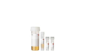
Human Aortic Endothelial Cells (HAoEC) are isolated from the human ascending (thoracic) and descending (abdominal) aorta. They are useful for studying vascular diseases such as thrombosis, atherosclerosis, and hypertension as well as for stent-graft compatibility testing.
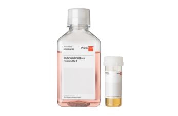
At PromoCell we guarantee for our primary human cells ≥ 500;000 viable cells after thawing. For this; we dispense > 500;000 cells per cryovial before cryopreservation as there will always be a certain percentage of dead cells after freeze/thaw.
In order to know the number of cells that survived the procedure; we defrost a representative number of vials per lot during QC; determine the cell viability using an electronic counting device and then calculate the number of viable cells that can be recovered after thawing. Both numbers - the calculated number of viable cells and the viability - can be found on the lot-specific Certificate of Analysis (CoA) that can be downloaded from our website.
Example: When the CoA indicates 600;000 viable cells and a viability of 80%; this means that the vial actually contains 750;000 cells (viable + dead); 80% thereof (600;000) were viable after thawing in our QC. We do not indicate the total number of cells per vial but just the number of expected viable cells which can be recovered when the recommended thawing protocol is used. You don't have to calculate any viabilities by yourself.
When the recommended plating density for your cell type is 5;000 - 10;000 cells/cm²; then the 600;000 viable cells can be plated e.g. in a T75 (corresponding to 8;000 cells/cm²) or in a T75 + a T25 (corresponding to 6;000 cells/cm²).
Some of our customers have successfully used PromoCell Endothelial Cell Growth Medium MV2 (C-22022) when co-culturing Human Coronary Artery Endothelial Cells (HCAEC; C-12221) and Normal Human Dermal Fibroblasts (NHDF; C-12300).
Our customers have successfully used TC flasks and dishes from all the leading cell culture plastic suppliers to grow PromoCell's primary human cells. We do not have any knowledge whether the dishes from local TC plastic suppliers work in the same way. We recommend to first test whether these brands provide the same good performance as the plastic of the leading manufacturers.
The time needed to detach our primary cells depends on many different factors like the cell type; cell density; lot #; trypsin concentration; the efficiency of the washing step before adding the trypsin and the trypsinization temperature.
For most cell types we recommend trypsinization at room temperature and direct observation of detachment under the microscope. This way; you can find out your individual trypsinization time and keep the contact time between cells and trypsin to a minimum. Most cells detach after 2-8 min.
Please refer to the instructions in the Manual. For some cell types; trypsinization at 37°C or the use of Accutase or another Detachment Solution is recommended.
Short protocol:
Trypsinize the cells as usual
Centrifuge and resuspend in suitable cold freezing medium at a density of 1-4 x 10⁶ cells/ml
Cool down the cells slowly to -80°C (approx. -1°C per min). We recommend to use "Mr. Frosty" from Nalge or "CoolCell" from Biocision; which both provide gradual and controlled cooling rates when placed in a -80°C freezer overnight.
Transfer the vials into liquid nitrogen for long-term storage
Our Normal Human Cells have been cultured and tested in our growth media and have adapted to these conditions. Using other media may yield unsatisfactory results due to suboptimal supplies of nutrients and growth factors. PromoCell can only guarantee good cell growth (as stated in the Certificate of Analysis) when the cells are grown in the recommended media.
PromoCell Growth Media have special formulations and are much more complex than DMEM or RPMI + FBS. In comparison to immortalized cell lines primary cells have much higher nutrient and growth factor requirements. Classical media do not usually achieve good performances with our cells.
It is not necessary to use coated flasks for (most of) our Normal Human Cells but it can be done. As coating with extracellular matrix proteins can affect cellular metabolism; it is recommended to use the same coating material for a complete set of experiments.
Cells that need to be grown on coated dishes:
Mesenchymal Stem Cells (C-12974/C-12971/C-12977) need Fibronectin-coating when grown in PromoCell MSC Growth Medium XF (C-28019) and when differentiated in MSC Neurogenic (C-28015); Adipogenic (C-28016); or Osteogenic (C-28013) Differentiation Media.
Human monocyte-derived macrophages (C-12914/C-12916/C-12915/C-12917) must be seeded into Fibronectin-coated culture vessels in combination with PromoCell's M1- and M2-Generation Media XF (C-28055; C-28056).
For efficient induction of osteoblast mineralization with PromoCell's Osteoblast Mineralization Medium (C-27020); the TC plates should be pre-coated with collagen type I.
The population doubling time or generation time (tg) is usually calculated during the logarithmic phase of growth. It specifies the time (t) in hours needed by the culture to double its cell number.
tg = t / n n: number of population doublings
Yes but the experimental starvation conditions have to be determined individually for each cell type. Usually the cells are maintained in basal medium with reduced growth factor concentrations or lower FBS content. The cells need to be in a good condition and the starvation should be kept as short as possible as prolonged serum and/or growth factor deprivation induces apoptosis.
Cell type-specific markers are usually determined one passage after thawing.
Example: After thawing; HUVEC are in P1. Markers (CD31 expression; Dil-Ac-LDL uptake) are tested in P2.
Please note: Cells that are frozen directly after isolation (blood cells) are tested directly after thawing with no further culturing step.
The passage number varies depending on the cell type. Please refer to the Manual for your cells under "Specifications".
You will also find the information in the Certificate of Analysis (CoA).
The recommended plating density after thawing/subculture may vary depending on the cell type. Please refer to the data sheet for your cells under "Specifications".
The Certificates of Analysis can easily be downloaded from our PromoCell website:
https://www.promocell.com/certificate-of-analysis/
Simply type in the lot number indicated on the cryovial/TC-flask and click the SEARCH button.
In principle; both types of liquid nitrogen storage are acceptable; each having its advantages and disadvantages.
Liquid phase storage provides a consistent temperature of -196°C; a longer holding time and a greater vial capacity but involves the risk of contamination issues.
Storage in the gas phase is very safe with respect to contaminations but the holding time of the cells is shorter and the vial capacity is reduced.
PromoCell guarantee 15 population doublings (PD) for most Normal Human Cells (unless otherwise indicated on the Certificate of Analysis) when the recommended PromoCell media and the PromoCell DetachKit are used. For more information; please check the respective Manual; section "Specifications".
PromoCell does not determine the number of passages but instead we calculate the population doublings (PD) that can be performed with the cells. The term passage only describes the process of detachment and replating and does not take into account different split ratios. The optimal split ratio is calculated from the actual cell yield after trypsinisation and the recommended plating density. In most of our cell types; the split ratio is usually between 1:3 and 1:6. Using 1:4 splits (i.e. increasing the growth surface by factor 4 each time); 15 doublings are achieved after 6-8 passages.
For recommended plating densities; please view the respective Manual; section "Specifications".
Freshly isolated cells that are plated in a tissue culture vessel for the first time are named primary cells or primary culture (corresponding to P0). As soon as they have been subcultured; they should correctly be termed normal cells (> P1).
Generally; the medium should be changed every 2-3 days.
Please note: Following thawing; the first medium change should be performed after 16-24 hours to prevent cell damage due to residual freezing medium.
General protocol for recovery of anchorage-dependent primary cells: Remove vial from liquid nitrogen; transport it on dry ice to the cell culture lab. Thaw in a 37°C waterbath for approx. 2 min; until it is just defrosted. Keep the vial immersed in water until just below the screw cap during thawing and only remove it shortly after approx. 90 sec to check the progress. Do not repeatedly insert and remove the vial while thawing in water. Carefully disinfect the vial with plenty of 70% EtOH under the laminar flow hood and aseptically transfer the thawed suspension into an appropriate TC dish with growth medium (pre-warmed in the incubator for > 30 min). The cells usually attach within a few hours. Perform media change after 24 hrs at the latest to remove residual DMSO from the freezing media. Additional information can be found in the instruction manuals of our cells.
Your observation can be either ascribed to a change of the culture medium to a cross-contamination with another cell type or to differentiation or senescence. The attached trouble shooting guide should help you to identify the reason for your observation.
Microbiological contaminations can be introduced into cell cultures by unsterile working techniques contaminated water baths media plasticware etc. Please check the attached trouble shooting guide for more detailed information.
Normal cells have a finite life span and therefore eventually stop growing and become senescent. In addition a cease in proliferation or cell death can be induced by other factors. For more details check the trouble shooting guide below.
Slow growth after subculture can be caused by over-trypsinization or other suboptimal culture conditions. Please see attached trouble shooting guide for possible reasons.
Several different methods for the detection of mycoplasmas have been described like e.g. cultures on agar in liquid or semi-solid media staining with DAPI mycoplasma-specific antibodies biochemical methods and PCR-based assays. PCR-based detection is very sensitive detects all mycoplasma species that occur in cell cultures and is completed within 3-5 hours.
The study of angiogenesis has been significantly advanced by the ability to culture endothelial cells in vitro. Initially large vessel ECs such as those isolated from the human umbilical vein (HUVEC) were used for these studies but increasingly it has been recognized that microvascular endothelial cells are a more appropriate model since angiogenesis involves microvessels rather than large vessel ECs.
There are several factors that can influence successful transfections; e.g. viability and density of the cells; choice of the transfection reagent; quality and type of the transfected molecules (plasmids; siRNA; oligonucleotides); as well as the culture medium and supplements used.
When using PromoCell Endothelial Cell Growth Media for cell transfection; please follow the instructions below:
Heparin; which is included in our Endothelial Cell Growth Medium (C-22010/C-22110); Medium 2 (C-22011/C-22111) and Medium MV (C-22020/C-22120) may reduce the transfection efficiency. We therefore recommend to use our heparin-free Endothelial Cell Growth Medium MV2 (C-22022/C-22121). Alternatively; you may use Endothelial Cell Growth Medium Kit (C-22110); Medium 2 Kit (C-22111); or Medium MV Kit (C-22120) without adding the ECGS/heparin supplement to the Basal Medium.
Please note: Before and after transfection; the cells should be cultured in complete Growth Medium including heparin to ensure optimal growth.
Secretory granules called Weibel-Palade bodies (WPBs) containing Von Willebrand factor (vWF) are more linked to the formation of a confluent endothelial monolayer. Research shows that vWF expression is dynamic and highly dependent on the cell culture conditions such as confluence and passage number.
Therefore; having vWF as a quality control marker for each lot of ECs is not really necessary.
We guarantee > 500;000 viable cells per vial. To do this; we need to freeze more than 500;000 cells; as we do not yet know the viability after thawing at the time of freezing.
For technical/organizational reasons; the initial cell number may also vary from lot to lot; so 2 lots with the same viability may not necessarily contain the same number of cells.
Based on negative feedback we have received from customers using bead baths; we strongly discourage the use of bead baths to thaw our cells. It can lead to reduced viability or significantly slower growing cells.
If you don’t have a “normal” water bath but only a bead bath in your lab; thaw the vial in a beaker of water in the bead bath. Ensure the water is heated to exactly 37°C using a thermometer placed in the warmed water. Be sure to hold the vial in your hand; and not in a floater; as described in the thawing protocol.
Our cells are not manufactured according to GMP guidelines and are intended for in vitro use only.
Our EXCiPACT™ GMP certification only applies to the processes related to the media.
The human leukocyte antigen (HLA) complex - relevance of HLA to cancer immunotherapy
HLA-Typed Primary Cells: The advantages of working with HLA in drug development research
Human cardiovascular cell types
HLA-typed human primary cells
Cell disease models
Recommended media for endothelial cells
Endothelial cells
HLA-typed human primary cells
The importance of HLA for the development of cell therapies
PromoCell uses the Bioz AI engine to display scientific citations for this product. This content is currently blocked because you haven’t enabled functional cookies.
To see this content, please click the cookie settings icon (bottom left corner of the screen) and enable functional cookies.
By clicking "Load content now", you agree to load content from Bioz, a third-party provider. This sets cookies on your device, without changing your saved cookie preferences.
Recommended products
Endothelial Cell Growth Medium MV
Low-serum cell culture medium for endothelial cells from microvascular vessels, the coronary artery, and the aorta.