


Technical library
Items 1-12 of 285 Results
 PromoCell_Best-Practices-in-Cell-Culture
tools-for-respiratory-research_brochure
Standardized culture of assay-ready and fully functional human primary macrophages
Human Mononuclear Cells from Peripheral Blood (hMNC-PB), single donor, ultra-pure
PromoCell_Best-Practices-in-Cell-Culture
tools-for-respiratory-research_brochure
Standardized culture of assay-ready and fully functional human primary macrophages
Human Mononuclear Cells from Peripheral Blood (hMNC-PB), single donor, ultra-pure
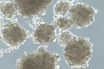
During the isolation of our Human Mononuclear Cells (hMNC), we first of all pay attention to thoroughly discard the platelet-containing fraction before we aspirate the MNC containing-interphase of the Ficoll gradient. The harvested MNC fraction is then subjected to several washing steps to remove potential remaining platelets. Finally, the cell preparations are verified by microscopy to be largely free of platelet contamination.
PromoCell_Best-Practices-in-Cell-Culture
tools-for-respiratory-research_brochure
Standardized culture of assay-ready and fully functional human primary macrophages
Human Mononuclear Cells from Peripheral Blood (hMNC-PB), single donor, ultra-pure
There is a strong variation from donor to donor and sometimes from donation to donation concerning the number of vials that can be produced. This is due to individual variances between the donors as some of them have more MNCs per ml of blood and others have less. Generally, the number of vials (each with 25 x 106 cryopreserved cells) per lot ranges between 10 and 40.
PromoCell_Best-Practices-in-Cell-Culture
tools-for-respiratory-research_brochure
Standardized culture of assay-ready and fully functional human primary macrophages
Choosing a suitable culture medium is crucial factor for in vitro cell cultivation and significantly affects the success of cell culture experiments, from the first step of development and when transitioning to clinical applications. Due to specific requirements of primary cells and each researcher’s application, we provide a wide range of advanced media formulations.
Therefore, it is essential for researchers to have a clear understanding of how to define the specifications of our cell culture media and reagents, from serum-free or xeno-free to chemically defined, to estimate and understand the associated implications for their intended applications. We can support you by providing a comprehensive guideline for the characterization and qualification of our cell culture media and reagents.
Discover and download our Media & Reagents Specification Guide
PromoCell_Best-Practices-in-Cell-Culture
tools-for-respiratory-research_brochure
Standardized culture of assay-ready and fully functional human primary macrophages
Human M1 Macrophages (GM-CSF), Monocyte-derived, single donor, 1,5 Mio cells
Human M2 Macrophages (M-CSF), Monocyte-derived, single donor, 1,5 Mio cells
M1-Macrophage Generation Medium XF
M2-Macrophage Generation Medium XF
Macrophage Base Medium XF
Without cytokines, the macrophages will die very quickly. You must supplement the Macrophage Base Medium XF (Bsal Medium + SupplementMix) with the appropriate cytokines or at least with human AB serum.
PromoCell_Best-Practices-in-Cell-Culture
tools-for-respiratory-research_brochure
Standardized culture of assay-ready and fully functional human primary macrophages
Primary Cancer Culture System
Our Primary Cancer Culture System (PCCS, C-28081) supports Cancer Stem Cells universally, therefore the PCCS can also be used for CSCs from blood cancer/leukaemia.
However, we recommend not to treat the surface of the culture vessel with NCCD reagent, but to add 12.5 µl NCCD/ml to the PCCS and seed the cells into the medium.
PromoCell_Best-Practices-in-Cell-Culture
tools-for-respiratory-research_brochure
Standardized culture of assay-ready and fully functional human primary macrophages
3D Tumosphere Medium XF, phenol red-free
3D Tumosphere Medium XF
Cancer Cell Line Medium XF
Primary Cancer Culture System
Yes, our cancer media (Primary Cancer Culture System/PCCS, 3D Tumorsphere Medium XF, Cancer Cell Line Medium XF) also support the growth of murine tumor cells.
PromoCell_Best-Practices-in-Cell-Culture
tools-for-respiratory-research_brochure
Standardized culture of assay-ready and fully functional human primary macrophages
Primary Cancer Culture System
Yes, it is. The Primary Cancer Culture System (C-28081) is very selective for cancer stem cells – any other cells will be eliminated after a short time.
App Note: Isolation of patient-derived cancer stem cells
PromoCell_Best-Practices-in-Cell-Culture
tools-for-respiratory-research_brochure
Standardized culture of assay-ready and fully functional human primary macrophages
Primary Cancer Culture System
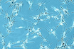
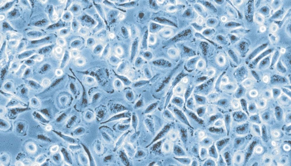
In our Primary Cancer Culture System (PCCS, C-28081), only the malignant tumor cells can survive. Therefore, benign animal cells (e.g., fibroblasts) will be depleted and the malignant human tumor cells will survive.
PromoCell_Best-Practices-in-Cell-Culture
tools-for-respiratory-research_brochure
Standardized culture of assay-ready and fully functional human primary macrophages
Human Chondrocytes (HCH)
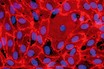
Alcian Blue stains the extracellular matrix of chondrocytes, e.g. cartilage-specific aggrecan and other glycosaminoglycans. To our knowledge, chondrocytes only express aggrecans when grown in 3-D culture and not in 2-D culture. To detect cartilage specific markers in monolayer culture, it is recommended to perform immunofluorescence detection of collagen type II.
PromoCell_Best-Practices-in-Cell-Culture
tools-for-respiratory-research_brochure
Standardized culture of assay-ready and fully functional human primary macrophages
Our skeletal muscle cells are mainly isolated from M. pectoralis, sometimes also from M. gastrocnemius, M. intercostales or M. gluteus maximus. The exact localization is specified in the Certificate of Analysis.
PromoCell_Best-Practices-in-Cell-Culture
tools-for-respiratory-research_brochure
Standardized culture of assay-ready and fully functional human primary macrophages
Trypsin Neutralizing Solution - 30 ml
Trypsin Neutralizing Solution - 125 ml
Trypsin Neutralizing Solution - 250 ml
Our TNS solution contains 0.05% (w/v) trypsin inhibitor from soy bean in HepesBSS/0.1% BSA.
PromoCell_Best-Practices-in-Cell-Culture
tools-for-respiratory-research_brochure
Standardized culture of assay-ready and fully functional human primary macrophages
Adipocyte Nutrition Medium (Ready-to-use)
Adipocyte Basal Medium
Airway Epithelial Cell Growth Medium (Ready-to-use)
Chondrocyte Basal Medium
Chondrocyte Growth Medium (Ready-to-use)
DC Base Medium
DC Generation Medium (Ready-to-use)
DC Base Medium XF
DC Generation Medium XF (Ready-to-use)
Endothelial Cell Growth Medium (Ready-to-use)
Endothelial Cell Growth Medium 2 (Ready-to-use)
Endothelial Cell Growth Medium MV (Ready-to-use)
Endothelial Cell Growth Medium MV2 (Ready-to-use)
Fibroblast Growth Medium (Ready-to-use)
Fibroblast Growth Medium 2 (Ready-to-use)
Fibroblast Growth Medium 3 (Ready-to-use)
Follicle Dermal Papilla Cell Growth Medium (Ready-to-use)
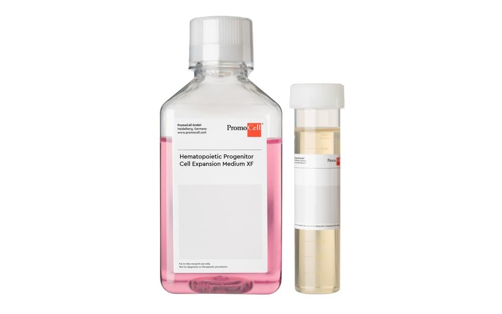
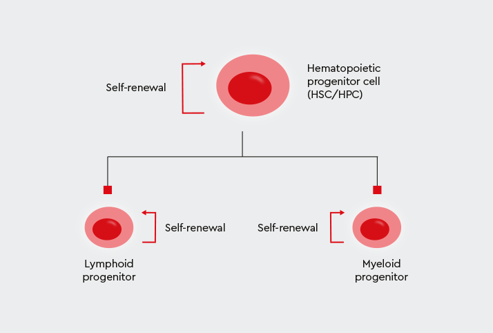
Hematopoietic Progenitor Cell Expansion Medium XF
Keratinocyte Growth Medium 2 (Ready-to-use)
Keratinocyte Growth Medium 3
M1-Macrophage Generation Medium XF
M2-Macrophage Generation Medium XF
Macrophage Base Medium XF
Mammary Epithelial Cell Growth Medium (Ready-to-use)
Melanocyte Growth Medium (Ready-to-use)
Mesenchymal Stem Cell Adipogenic Differentiation Medium 2
Mesenchymal Stem Cell Chondrogenic Differentiation Medium - 100, ml
Mesenchymal Stem Cell Chondrogenic Differentiation Medium - 100, ml
Mesenchymal Stem Cell Growth Medium 2
Mesenchymal Stem Cell Neurogenic Differentiation Medium
Mesenchymal Stem Cell Osteogenic Differentiation Medium
Monocyte Attachment Medium
Mononuclear Cell Medium
Myocyte Growth Medium (Ready-to-use)
Osteoblast Growth Medium (Ready-to-use)
Osteoblast Mineralization Medium
Preadipocyte Growth Medium (Ready-to-use)
Renal Epithelial Cell Growth Medium 2 (Ready-to-use)
Skeletal Muscle Cell Growth Medium (Ready-to-use)
Skeletal Muscle Cell Differentiation Medium (Ready-to-use)
Small Airway Epithelial Cell Growth Medium (Ready-to-use)
Smooth Muscle Cell Growth Medium 2 (Ready-to-use)
Mesenchymal Stem Cell Growth Medium XF
Melanocyte Growth Medium M3 (Ready-to-use)
Yes, you can aliquot the SupplementMix upon delivery and freeze down 2 or 4 individual aliquots at -20°C. This way, you can prepare smaller volumes (2 x 250 ml or 4 x 125 ml) of complete culture medium and thus extend the time you can use the medium.