


Items 1-12 of 20 Results
-

The immunomodulatory properties of mesenchymal stem cells (MSCs)
Since their discovery in 1991, mesenchymal stem cells, or MSCs, have been used as a promising method to build tissue. However, MSCs can do more for the development of cell and gene therapies. Find out more in our video. Video
Video
-

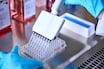
Thawing frozen cells – detailed protocol for human primary cells
Thawing cells correctly is absolutely necessary when it comes to successfully starting reliable cell cultures. Find out how to thaw frozen primary cells properly in our detailed video protocol.
Video
-
Primary cells in respiratory research – webinar
Learn about the benefits of primary cells as model systems in respiratory research and how a 3D model at the Air-Liquid Interface can be set up and used.
Video
-
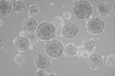
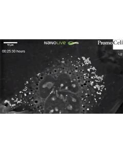
Cells in Action: Mitosis in Human Mesenchymal Stem Cells (MSCs)
Watch the process of cell division in PromoCell's human mesenchymal stem cells! These images were captured using Nanolive’s non-invasive technology, which allows us to access the dynamics of biological processes, such as mitosis, with unprecedented resolution.
Video
-
How to achieve a reliable Air-Liquid Interface (ALI) Culture
Watch our explainer video to find out how to use our Air-Liquid Interface Culture system for respiratory research in an environment that replicates in vivo.
Video
-
Improve the reproducibility of your research with standardized primary cell culture techniques
This webinar provides expert’s tips and tricks in successfully handling human primary cells and learn how to standardize primary cell culture techniques.
Video
-

HLA-Typed Primary Cells: The advantages of working with HLA in drug development research
For those working in drug development, human leukocyte antigens or HLA are a key element to keep in mind as an essential regulator of the immune system. Watch our latest explainer video to understand the benefits of working with HLA in your pre-clinical research.
Video
-
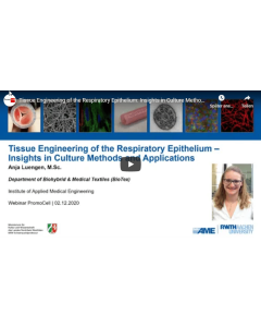
Tissue engineering of the respiratory epithelium: insights in culture methods and applications
This webinar addresses both interested beginners as well as advanced scientists in the field and focuses on key issues like cell sources, air liquid interface (ALI) culture, medium demands and current challenges regarding reproducibility and standardization.
Video
-
Getting started with primary cells – Standard procedures in cell culture
In this quick start guide, we review the finer points of thawing, plating, and passaging primary human cells. Even if you’re experienced using these cells, we review some of the little things that can make a large difference in your cell culture success with our cells. Presented by PromoCell Application Specialist, Dr Melissa Olekson.
Video
-
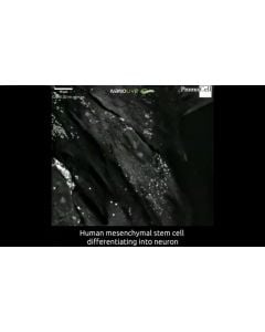
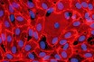
Cells in action: Differentiation of human mesenchymal stem cells into neurons
Cells in action: Differentiation of human mesenchymal stem cells into neurons In this video we can see the differentiation of PromoCell umbilical cord mesenchymal stem cells into neurons. Excitingly, to our knowledge this is one of the first high-resolution, long-term, live cell time-lapses showing this process. These cells were grown for 13 days in PromoCell complete mesenchymal stem cell neurogenic differentiation medium prior to capture of this film. Images were captured every 30 seconds over a 20-hour period using NanoLive’s 3D Cell Explorer.
Video
-

MSC reproducibility: Towards the standardization of Mesenchymal Stem Cells
For science to move forwards, the research we do must be reproducible. One of the ways we’re helping your work be as consistent as possible is through our characterized MSCs.
Video
-
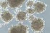
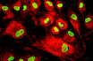
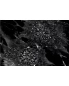
Cells in Action: Human Primary Keratinocytes at the stratum granulosum
Keratinocytes at the stratum granulosum contain two characteristic structures that are visible in this high resolution footage by Nanolive - the lamellar bodies and keratohyalin granules. While keratohyalin granules contain proteins involved in the aggregation of keratin filaments and in the formation of the cell envelope, lamellar bodies contain lipids and fuse with the plasma membrane in order to secrete the content into the extracellular space.
Video