The wound healing assay, also commonly referred as “scratch assay”, is a widely established technique to study collective cell migration in vitro. In a collaborative effort between CytoSMART and PromoCell, we demonstrate how this assay can be easily performed with primary human keratinocytes using software-based data analysis to achieve more accurate results.
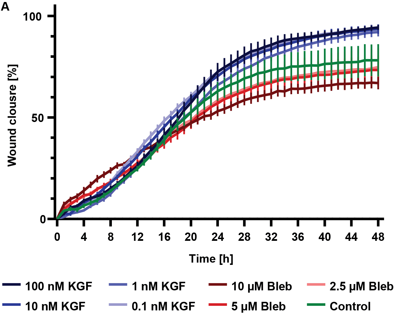
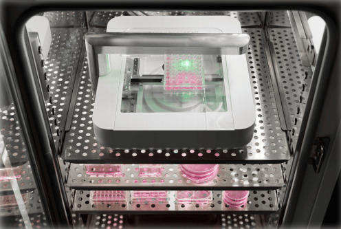
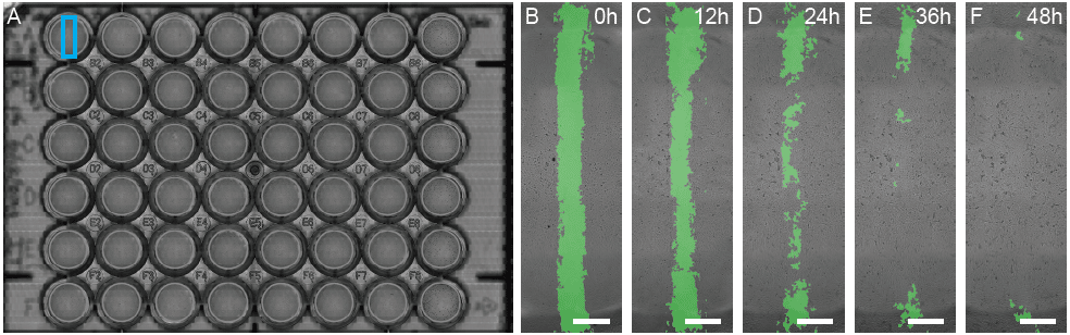
In this experiment, the CytoSMART Omni was used to analyze the migratory activity of human primary dermal keratinocytes in wound healing experiments by live-cell imaging. In combination with the included software analysis algorithm, it is possible to quantify the gap surface closure and closure speed over time. Acquiring data with a time-lapse microscope offers the advantage of documenting the wound assay under defined and stable conditions inside an incubator.
Typically, the cell types used in a wound healing assay aim to recreate the regenerative features of the epidermis. Cell lines as well as primary cells are often used as a model for this. The most commonly used cell types are Keratinocytes and Fibroblasts. In contrast to cell lines, human primary cells offer the advantage of allowing to investigate donor-dependent effects such as as age, sex or known disease conditions. In addition, they are closer representation of the in vivo environment as they are not sourced from malignant tissue, nor have been adapted to cell culture conditions over a large number of passages.
Commonly used primary human dermal cell types in wound healing assays: