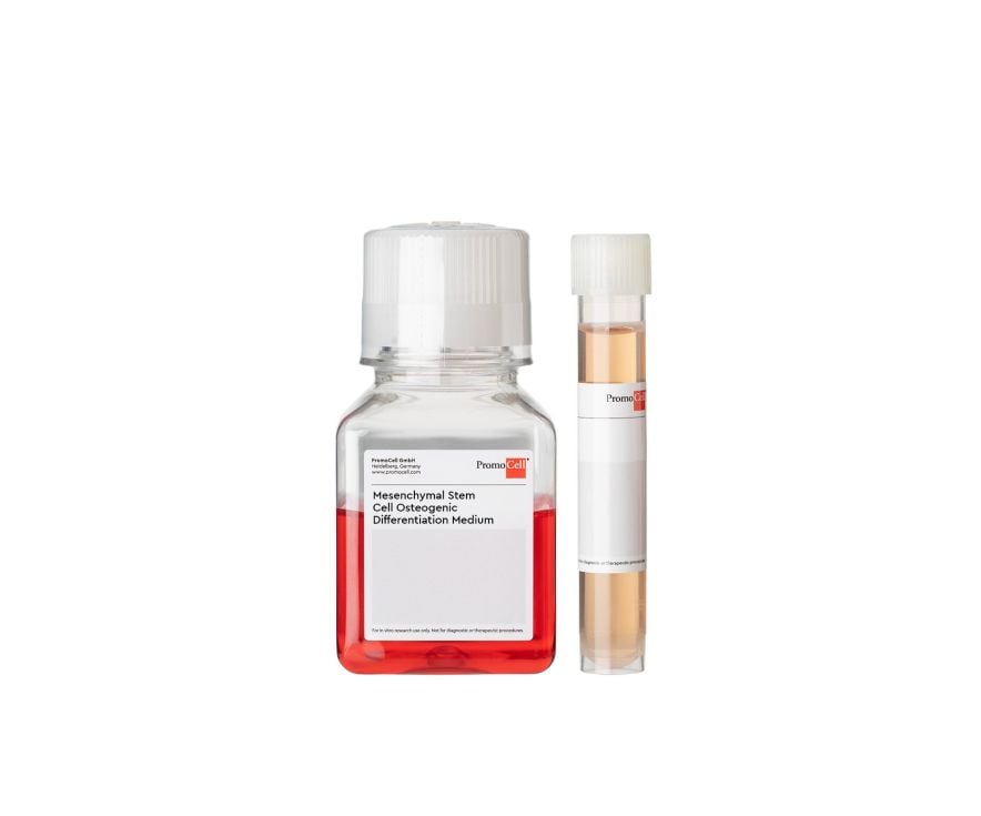
Mesenchymal Stem Cell Osteogenic Differentiation Medium
 Powered by Bioz
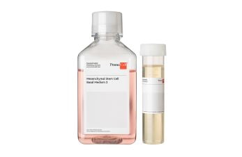
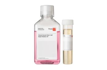
We offer a complete Mesenchymal Stem Cell Media System including growth media, differentiation media, and human mesenchymal stem cells (MSC).
We offer five MSC Differentiation Media to efficiently induce differentiation of MSC into adipogenic, chondrogenic (with and without inducers), osteogenic or neurogenic lineages, respectively.
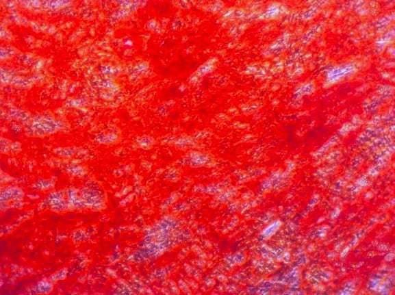
Our Mesenchymal Stem Cell Osteogenic Differentiation Media was developed for the directed differentiation of Human Mesenchymal Stem Cells (MSC) from bone marrow, the umbilical cord matrix (Wharton's Jelly) and adipose tissue into osteogenic lineages.
Although all our media are optimized for use with primary human cells, we have received feedback from customers that this particular medium can also be used for porcine cells.
View a list of references where this medium was used with other species and cell types.
PromoCell uses the Bioz AI engine to display scientific references for this product. This content is currently blocked because functional cookies are disabled.
Interested in our scientific references?
Click on the following link to load the content or enable functional cookies in the consent settings.
By clicking "Load content now", you agree to load content from Bioz, a third-party provider. This will set cookies on your device without changing your saved cookie preferences.