Human preadipocyte cell models are widely used to study adipogenesis, obesity, insulin resistance, and metabolic disease mechanisms in vitro. However, assay outcomes can vary depending on donor background, adipose depot origin, differentiation capacity, and culture conditions. Selecting the appropriate human preadipocyte model is therefore essential for improving reproducibility, translational relevance, and metabolic assay consistency.
In this article, we break down the key differences between available human adipocyte in vitro models, explain what drives assay variability, and give you practical tools for improving the reproducibility of your experiments.
Quick answers: Which human preadipocyte model should you choose?
When should I choose primary human preadipocytes?
Choose primary human preadipocytes when donor background, adipose depot, disease relevance, or translational metabolic responses are important.When should I choose immortalized adipocyte cell lines?
Choose immortalized adipocyte cell lines when scalability, simplified handling, or early-stage screening throughput are the main priorities.When should I consider adipose-derived stem cells?
Consider adipose-derived stem cells when the study focuses on progenitor cell biology, lineage commitment, or patient-specific differentiation.How can I improve adipocyte assay reproducibility?
Improve adipocyte assay reproducibility by standardizing passage number, seeding density, media conditions, differentiation timing, and assay endpoints.Why does adipocyte model selection matter in metabolic disease research?
The human preadipocyte model you choose can influence what your assay reveals and how confidently results can be interpreted. It can affect differentiation efficiency, insulin responsiveness, lipid accumulation, inflammatory signaling, glucose uptake, and drug sensitivity. These variables shape both the reproducibility and translational relevance of metabolic assays.1,2
For research into obesity, type 2 diabetes, or insulin resistance, this matters. To generate meaningful data, your model needs to reflect human adipose biology as closely as possible. Animal models have been widely used, but they fail to capture species-specific differences in adipocyte metabolism and depot-specific responses.3
Human preadipocyte cell models can help bridge this gap by providing human-relevant systems for adipogenesis and metabolic disease research. But not all human cell models are the same. Primary human preadipocytes, immortalized adipocyte cell lines, and adipose-derived stem cell systems have different properties and behaviors. Using the wrong model for your assay type can introduce variability, reduce translational relevance, or make results difficult to reproduce.4
In short, the more closely your model matches your research question, the more reliable and actionable your data becomes.
For a broad overview of adipocyte biology and metabolic function, read our foundational article on the role of adipocytes in metabolic homeostasis.
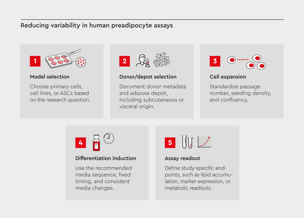
Figure 1. Key variables influencing adipocyte differentiation assay reproducibility in human preadipocyte models.
Human preadipocyte assay outcomes are shaped by both biological and technical variables. Model selection, donor and depot characteristics, cell expansion conditions, differentiation induction, and assay readout timing can all influence adipocyte differentiation and downstream metabolic readouts. Documenting biological variability while standardizing technical factors such as passage number, seeding density, media conditions, and endpoint timing can help improve consistency across adipocyte differentiation assays.
What types of human adipocyte models are available?
Three main model categories exist for in vitro adipogenesis research. Each model type supports different experimental goals and comes with specific strengths and limitations.
| Model type | Primary human preadipocytes | Immortalized cell lines | Adipose-derived stem cells |
|---|---|---|---|
| Biological relevance | High; reflects human physiology | Moderate; may lack key metabolic responses | High; patient-derived, depot-specific |
| Donor variability | Present; managed by donor selection | Low; clonal or pooled cell lines | Present; donor-dependent |
| Differentiation consistency | High with standardized protocols | High; predictable behavior | Moderate; requires optimization |
| Scalability | Moderate; limited by donor supply | High; easy to expand | Moderate |
| Translational relevance | High; suitable for disease modeling | Lower; may not reflect in vivo responses | High; depot-specific studies |
| Typical applications | Disease modeling, drug screening, metabolic assays | High-throughput screening, mechanistic studies | Depot comparison, patient-specific research |
Table 1. Comparison of human preadipocyte and adipocyte in vitro model types across key experimental criteria.4,5
Primary human preadipocytes
Use primary human preadipocytes when physiological relevance and donor-specific metabolic behavior are important experimental variables.
These cells are isolated directly from human adipose tissue, including subcutaneous and visceral depots. They retain donor-specific biology such as metabolic and inflammatory characteristics.6 This makes them a physiologically relevant option for disease modeling and selected drug screening applications.
For studies that depend on donor- and tissue-relevant biology, primary human preadipocytes can provide important biological context for interpreting metabolic assay results.
Immortalized adipocyte cell lines
Immortalized adipocyte cell lines are useful when consistency, scalability, simplified handling, or early-stage screening throughput are the main priorities.
They are genetically immortalized cells and therefore offer:
- predictable behavior
- easy expansion
- low batch-to-batch variability
However, immortalized adipocyte cell lines may show altered differentiation behavior, reduced depot specificity, and weaker representation of donor-specific metabolic phenotypes compared with primary cells.4,7
For studies that prioritize throughput or simplified assay setup, immortalized cell lines can offer practical advantages. For donor- or depot-specific metabolic questions, primary human models may provide more relevant biological context.
Adipose-derived stem cells (ASCs)
ASCs are primary multipotent cells and can be directed toward adipogenic differentiation. They can be useful when the study focuses on progenitor cell biology, lineage commitment, depot-specific research, or patient-specific differentiation.8,9
For studies that require a broader progenitor-cell context, ASCs can support adipogenic differentiation workflows while allowing researchers to investigate earlier stages of adipocyte lineage development.
What is the difference between primary preadipocytes and adipocyte cell lines?
Choosing between primary preadipocytes and adipocyte cell lines starts with a practical question: does your assay need donor-relevant biology, experimental scalability, or both?
Primary human preadipocytes are often more suitable when donor background, adipose depot, disease state, or metabolic response are central to the research question.
Meanwhile, immortalized adipocyte cell lines may be more suitable when consistency, scalability, simplified handling, or high-throughput screening are the main priorities.
For translational metabolic assays, donor-relevant primary human preadipocyte models can provide important biological context. For early-stage screening or simplified mechanistic studies, immortalized cell lines may offer practical advantages.
At PromoCell, we support your research workflows with donor- and depot-relevant primary human preadipocytes from defined adipose tissue origins along with corresponding media for expansion and differentiation.
Why do tissue origin and donor variability matter?
Even when using the same culture conditions and differentiation protocol, human preadipocytes can behave differently depending on the adipose depot and donor from which they were derived. Understanding these sources of biological variation can help guide model selection and improve interpretation of metabolic assay results.
Visceral vs subcutaneous preadipocytes: How does depot origin affect model selection?
Visceral and subcutaneous preadipocytes differ in inflammatory signaling, adipogenic behavior, and metabolic function, making depot selection an important experimental variable. With appropriate culture conditions, both visceral and subcutaneous preadipocytes can support adipogenic differentiation and develop hallmark features of adipocytes.
- Visceral preadipocytes show greater inflammatory cytokine secretion and stronger links to insulin resistance.11 This makes them particularly relevant for studying visceral adiposity, metabolic inflammation, and insulin resistance.
- Subcutaneous preadipocytes have a stronger lipid-storage phenotype.11,12 They are commonly used to study adipogenesis, lipid storage, and adipose tissue expansion.
In addition, lipolytic responses, adipokine profiles, inflammatory signaling, and sensitivity to glucocorticoids differ between the two depots.11
Depot-specific differences become especially important in research on obesity, metabolic syndrome, and insulin resistance. For example, white preadipocytes from subcutaneous adipose tissue have been used to explore adipogenesis, metabolism, and lipolysis.13-16
What this means for your research: The right adipose depot depends on the question your assay needs to answer. Selecting the appropriate depot helps align your human preadipocyte model with the biology you want to study.
How does donor variability affect adipocyte research?
Donor-specific variability is an important feature of primary human preadipocyte models. It can help reflect biologically relevant differences between donors and can be systematically addressed through experimental design, donor documentation, and standardized culture conditions.
Each donor brings a unique genetic background, including:
- age
- sex
- BMI
- metabolic status
These factors influence adipocyte differentiation, lipid accumulation, and responses to metabolic stimuli.17
Strategies that help address donor variability include using cells from matched donors across conditions, documenting donor metadata, pooling cells from multiple donors where appropriate, and maintaining consistent passage numbers.17
What causes variability in human adipocyte differentiation assays?
Variability in human adipocyte differentiation assays can arise from both biological and technical sources. Identifying where that variability comes from is the first step toward improving assay consistency.
Biological variability reflects meaningful differences between donors, depots, or disease states. Technical variability often results from differences in cell handling, culture conditions, media timing, or assay setup.
The following biological and technical variables are among the most common contributors to variability in human adipocyte differentiation assays.
Biological factors influencing adipocyte differentiation variability
Donor-to-donor differences, depot origin, disease state, and passage number can contribute to variation in adipogenic differentiation, lipid accumulation, and downstream metabolic readouts.
| Biological variable | Potential impact on assay outcomes |
|---|---|
| Donor-to-donor differences | Variable adipogenic differentiation capacity, lipid accumulation, and metabolic response |
| Depot origin | Depot-specific differences in adipogenic behavior, inflammatory signaling, lipid metabolism, and response to metabolic stimuli |
| Disease state | Altered differentiation behavior, lipid storage, adipokine secretion, or response to metabolic stimuli |
| Genetic background, age, sex, and BMI | Differences in baseline cell behavior, differentiation potential, and downstream metabolic readouts |
| Passage number | Changes in differentiation potential or cell behavior over extended culture |
Table 2: Biological variables influencing human adipocyte differentiation assays variability
Technical factors influencing adipocyte differentiation variability
Seeding density, confluency at induction, media handling, media-change timing, and assay endpoint definition can contribute to technical variability in human adipocyte differentiation assays.
| Technical variable | Potential impact on assay outcomes |
|---|---|
| Seeding density | Uneven cell growth and variable confluency before differentiation induction |
| Confluency at induction | Differences in adipogenic induction and reduced comparability between experiments |
| Media handling or non-standardized differentiation conditions | Inconsistent adipogenic induction and reduced comparability between experiments |
| Timing of media changes | Altered exposure to differentiation or nutrition conditions |
| Cell handling, including temperature shifts or mechanical stress | Reduced cell consistency before or during differentiation |
| Incubation time before assay readout | Variation in lipid accumulation, marker expression, or other study-specific endpoints |
| Assay endpoint definition | Reduced comparability of downstream metabolic readouts across experiments |
Table 3: Technical variables influencing human adipocyte differentiation assay variability.
Variability in adipogenesis assays can compromise reproducibility and reduce confidence in downstream metabolic readouts. Human adipocyte differentiation assays are particularly sensitive to changes in induction timing and media conditions.
Even small changes in culture conditions can influence differentiation outcomes and downstream readouts such as lipid accumulation, adipogenic marker expression, or glucose uptake. Depending on the study design, differentiated adipocytes may also be assessed using endpoints such as adipokine secretion, lipolysis, inflammatory cytokine response, or metabolic activity.18-20
How can standardized cells and matched media improve adipocyte assay reproducibility?
Standardized cells, suitable media, and defined protocols are key to reducing technical variability in human preadipocyte assays.21,22 While donor-specific variability is an important feature of primary cell models, researchers can improve reproducibility by controlling variables such as cell source, passage number, seeding density, media formulation, differentiation timing, and assay endpoint.
Start with the right cell model foundation
For primary human preadipocyte models, reproducibility begins with careful model selection. This includes choosing the appropriate adipose depot and documenting relevant donor characteristics, such as age, sex, BMI, and metabolic status, where available.23 This helps you interpret biological variation while reducing avoidable technical variability.
Improve workflow consistency with recommended cell and media combinations
Matched cell and media systems support workflow consistency. Low-serum growth media and defined, animal component-free differentiation media, for example, can help standardize the transition from preadipocyte expansion to adipocyte differentiation. This reduces the need for researchers to prepare complex differentiation formulations themselves.
Our human white preadipocyte differentiation application note provides a protocol example using human white preadipocytes from subcutaneous or visceral adipose tissue. The workflow includes defined timing for confluence, differentiation induction, regular media changes, and maturation into lipid-containing adipocytes.
How can researchers improve reproducibility in metabolic assays?
Reproducible adipocyte assays depend on standardized culture conditions, consistent differentiation timing, and careful donor documentation.24,25 The following workflow addresses the most common failure points.
Reproducibility checklist for human preadipocyte assays
- Model selection: Choose a human preadipocyte cell model that is suitable for your research question (depot, disease state, scalability).
- Donor documentation: Record donor age, sex, BMI, and passage number. Use early-passage cells where possible.
- Seeding standardization: Seed at a consistent density, and wait for full confluency before induction.
- Differentiation media: Use a standardized differentiation medium to reduce variability from media preparation and supplementation.
- Media change timing: Stick to your protocol’s schedule.
- Temperature and handling: Minimize time outside the incubator. Pre-warm reagents and avoid mechanical stress during pipetting.
- Assay timing: Define your assay endpoint precisely, as variation in endpoint timing can influence your results.
- Positive controls: Include an internal differentiation reference or positive control where appropriate.
How are human preadipocyte models advancing metabolic disease research?
Human preadipocyte models are used across metabolic disease research to study adipogenesis, adipose tissue dysfunction, insulin resistance, and disease-relevant metabolic responses in vitro. Depending on the research question, these models can support studies of obesity, type 2 diabetes, and metabolic syndrome.26
Different applications require different levels of model complexity, physiological relevance, reproducibility, and donor specificity. This makes model selection an important step when designing human-relevant metabolic assays.
Obesity and insulin resistance
Primary human preadipocyte cell models from donors with obesity or diabetes allow you to study disease mechanisms. Differences in differentiation capacity, lipid turnover, adipokine secretion, and response to metabolic stimuli between healthy and metabolically compromised donors can be investigated using primary human preadipocytes.6,27
Drug screening
Human-relevant screening models are increasingly important for metabolic disease research, where species differences and model-specific limitations can affect the interpretation of assay results. Primary human preadipocytes can provide donor- and depot-relevant context for studying responses to candidate compounds such as PPARγ agonists, insulin sensitizers, and lipogenic inhibitors.28,29
If you’re building metabolic disease screening workflows, our diabetes research solutions include primary human cell models, media, and protocols that support adipogenesis assays and functional metabolic readouts.
Inflammatory co-culture models
Co-culture systems that combine adipocytes with macrophages, endothelial cells, or immune cells are becoming an important tool for studying adipose tissue inflammation and cell-cell communication.30 For example, co-culture models with adipocytes and endothelial cells can help researchers investigate how adipose-derived signals may influence vascular function.31
Donor-specific and precision medicine applications
Donor-specific adipocyte models may support future precision medicine approaches by enabling patient-relevant metabolic phenotyping in vitro. As metabolic disease research becomes more focused on donor-specific variation, primary human preadipocyte models can help researchers investigate how donor background, disease state, and adipose depot may influence adipocyte differentiation and metabolic response.32
Choosing a human preadipocyte model starts with the research question
The optimal human preadipocyte model depends on whether the study prioritizes physiological relevance, donor specificity, scalability, or assay consistency. Primary human preadipocytes are particularly useful when donor background, adipose depot, and disease-relevant metabolic responses are central to the research question. Immortalized adipocyte cell lines may be more suitable when simplified handling, scalability, or high-throughput screening are the main priorities.
For reliable adipocyte differentiation assays, model selection should be combined with standardized culture conditions, suitable growth and differentiation media, and clearly defined assay endpoints. Aligning the model, protocol, and readout strategy helps researchers distinguish meaningful biological variation from avoidable technical variability and supports more reproducible metabolic data.
We provide primary human preadipocytes, low-serum growth media, and defined, animal component-free differentiation media to support human adipocyte in vitro workflows from cell expansion through adipocyte differentiation.
Building a human preadipocyte assay workflow? Model choice, donor background, adipose depot, media, and assay endpoints can all influence your results. If you need support selecting the right cells and media for your application, our scientific support team can help.
Frequently asked questions
References
Expand
- Todorčević M, Hilton C, McNeil C, et al. A cellular model for the investigation of depot specific human adipocyte biology. Adipocyte. 2017;6(1):40-55. doi:10.1080/21623945.2016.1277052
- Niesler CU, Siddle K, Prins JB. Human preadipocytes display a depot-specific susceptibility to apoptosis. Diabetes. 1998;47(8):1365-1368. doi:10.2337/diab.47.8.1365
- Börgeson E, Boucher J, Hagberg CE. Of mice and men: Pinpointing species differences in adipose tissue biology. Front Cell Dev Biol. 2022;10:1003118. doi:10.3389/fcell.2022.1003118
- Gutiérrez-García A, Olivas-Aguirre FJ, Olivas-Aguirre M. Critical evaluation of adipogenic cell models: Impact of the receptor toolkit on adipogenic potential. Receptors. 2025;4(4):19. doi:10.3390/receptors4040019
- Lauschke VM, Hagberg CE. Next-generation human adipose tissue culture methods. Curr Opin Genet Dev. 2023;80:102057. doi:10.1016/j.gde.2023.102057
- Ruiz-Ojeda F, Rupérez A, Gomez-Llorente C, Gil A, Aguilera C. Cell models and their application for studying adipogenic differentiation in relation to obesity: A review. Int J Mol Sci. 2016;17(7):1040. doi:10.3390/ijms17071040
- Wang QA, Scherer PE, Gupta RK. Improved methodologies for the study of adipose biology: Insights gained and opportunities ahead. J Lipid Res. 2014;55(4):605-624. doi:10.1194/jlr.R046441
- Tsuji W. Adipose-derived stem cells: Implications in tissue regeneration. World J Stem Cells. 2014;6(3):312. doi:10.4252/wjsc.v6.i3.312
- Chu DT, Nguyen Thi Phuong T, Tien NLB, et al. Adipose tissue stem cells for therapy: An update on the progress of isolation, culture, storage, and clinical application. J Clin Med. 2019;8(7):917. doi:10.3390/jcm8070917
- Bahmad HF, Daouk R, Azar J, et al. Modeling adipogenesis: Current and future perspective. Cells. 2020;9(10):2326. doi:10.3390/cells9102326
- Lin F, Sul HS. Distinct precursor landscape of subcutaneous and visceral fat in development and aging. Cell Rep. 2026;45(1):116706. doi:10.1016/j.celrep.2025.116706
- Li Y, Zhang H, Ibáñez CF, Xie M. Characterization of subcutaneous and visceral de-differentiated fat cells. Mol Metab. 2025;93:102105. doi:10.1016/j.molmet.2025.102105
- Ding C, Wilding JPH, Bing C. 1,25-dihydroxyvitamin D3 Protects against Macrophage-Induced Activation of NFκB and MAPK Signalling and Chemokine Release in Human Adipocytes. PLoS ONE. 2013;8(4):e61707. doi:10.1371/journal.pone.0061707
- Tourniaire F, Romier-Crouzet B, Lee JH, et al. Chemokine expression in inflamed adipose tissue is mainly mediated by NF-κB. PLoS ONE. 2013;8(6):e66515. doi:10.1371/journal.pone.0066515
- Zilleßen P, Celner J, Kretschmann A, Pfeifer A, Racké K, Mayer P. Metabolic role of dipeptidyl peptidase 4 (DPP4) in primary human (pre)adipocytes. Sci Rep. 2016;6(1):23074. doi:10.1038/srep23074
- Sancar G, Liu S, Gasser E, et al. FGF1 and insulin control lipolysis by convergent pathways. Cell Metab. 2022;34(1):171-183.e6. doi:10.1016/j.cmet.2021.12.004
- Beylerli O, Gareev I, Zhao B, Musaev E. Donor variability in adipose tissue-derived stem cells: Implications for the clinical efficacy of autologous fat grafting. Explor Med. 2024:601-614. doi:10.37349/emed.2024.00243
- Sheng X, Tucci J, Malvar J, Mittelman SD. Adipocyte differentiation is affected by media height above the cell layer. Int J Obes. 2014;38(2):315-320. doi:10.1038/ijo.2013.96
- Kraus NA, Ehebauer F, Zapp B, Rudolphi B, Kraus BJ, Kraus D. Quantitative assessment of adipocyte differentiation in cell culture. Adipocyte. 2016;5(4):351-358. doi:10.1080/21623945.2016.1240137
- Kudo T, Zhao ML, Jeknić S, et al. Context-dependent regulation of lipid accumulation in adipocytes by a HIF1α-PPARγ feedback network. Cell Syst. 2023;14(12):1074-1086.e7. doi:10.1016/j.cels.2023.10.010
- Panella S, Muoio F, Jossen V, Harder Y, Eibl-Schindler R, Tallone T. Chemically defined xeno- and serum-free cell culture medium to grow human adipose stem cells. Cells. 2021;10(2):466. doi:10.3390/cells10020466
- Regmi A, Roell W. Differentiation of human subcutaneous adipocytes and measurement of lipolytic function induced by GIP or LY3437943. STAR Protoc. 2023;4(2):102304. doi:10.1016/j.xpro.2023.102304
- Aranaz P, Clavel-Millan M, Gil-Cardoso K, et al. Preclinical research in obesity-associated metabolic diseases using in vitro, multicellular, and non-mammalian models. J Physiol Biochem. 2025;81(4):1225-1255. doi:10.1007/s13105-025-01130-6
- Kassotis CD, Masse L, Kim S, Schlezinger JJ, Webster TF, Stapleton HM. Characterization of adipogenic chemicals in three different cell culture systems: Implications for reproducibility based on cell source and handling. Sci Rep. 2017;7(1):42104. doi:10.1038/srep42104
- Ren XM, Chang RC, Amato AA, et al. Development and characterization of a standardized adipogenesis assay for testing metabolism disrupting chemicals using human bone marrow derived mesenchymal stem cells. NAM J. 2025;1:100029. doi:10.1016/j.namjnl.2025.100029
- Andersen E, Ingerslev LR, Fabre O, et al. Preadipocytes from obese humans with type 2 diabetes are epigenetically reprogrammed at genes controlling adipose tissue function. Int J Obes. 2019;43(2):306-318. doi:10.1038/s41366-018-0031-3
- Kowalczyk M, Piwowarski JP, Wardaszka A, Średnicka P, Wójcicki M, Juszczuk-Kubiak E. Application of in vitro models for studying the mechanisms underlying the obesogenic action of endocrine-disrupting chemicals (EDCs) as food contaminants—A review. Int J Mol Sci. 2023;24(2):1083. doi:10.3390/ijms24021083
- Chen Z, Yu H, Shi X, et al. Functional screening of candidate causal genes for insulin resistance in human preadipocytes and adipocytes. Circ Res. 2020;126(3):330-346. doi:10.1161/CIRCRESAHA.119.315246
- Sauma L, Stenkula KG, Kjølhede P, Strålfors P, Söderström M, Nystrom FH. PPAR-γ response element activity in intact primary human adipocytes: Effects of fatty acids. Nutrition. 2006;22(1):60-68. doi:10.1016/j.nut.2005.04.011
- Padmanaban AM, Ganesan K, Ramkumar KM. A co-culture system for studying cellular interactions in vascular disease. Bioengineering. 2024;11(11):1090. doi:10.3390/bioengineering11111090
- Brown OI, Bridge KI, Straw S, et al. Studying adipose endothelial cell/adipocyte cross-talk in human subcutaneous adipose tissue. J Vis Exp. 2024;(206):66608. doi:10.3791/66608
- Chami N, Wang Z, Svenstrup V, et al. Genetic subtyping of obesity reveals biological insights into the uncoupling of adiposity from its cardiometabolic comorbidities. Nat Med. 2025;31(11):3801-3812. doi:10.1038/s41591-025-03931-0
- Anatildes Da Silva De Paula GL, Correia Garcia E, Teles Soares Beserra B, Amorim Amato A. Thermogenic differentiation of human adipocyte precursors in culture: A systematic review. Cells. 2025;14(23):1907. doi:10.3390/cells14231907
- Lynes MD, Tseng Y. Deciphering adipose tissue heterogeneity. Ann N Y Acad Sci. 2018;1411(1):5-20. doi:10.1111/nyas.13398
- Ma X, Lee P, Chisholm DJ, James DE. Control of adipocyte differentiation in different fat depots; implications for pathophysiology or therapy. Front Endocrinol. 2015;6. doi:10.3389/fendo.2015.00001
Related resources