
Autoimmune disease rarely plays out in a single cell type. Dysregulated immune responses can affect specific tissues, while cells within those tissues can in turn shape local inflammation. Studying these mechanisms therefore often means looking beyond immune cells in isolation and considering the interactions between immune and tissue-specific cells.
Human primary cells offer a controlled way to investigate these processes in a human cellular setting and form part of the broader move toward human-relevant New Approach Methodologies (NAMs). In this article, we explore how primary-cell models can help represent immune–tissue interactions, when donor and HLA context matter, and how to select model components and readouts that fit your research question.
What are New Approach Methodologies (NAMs)?
New Approach Methodologies (NAMs) encompass a range of innovative approaches used to improve the human relevance of biomedical research and drug development. They include in vitro human-based systems, in silico models, and other emerging platforms.1
Their role in drug development is receiving increasing regulatory attention. FDA draft guidance issued in March 2026, for example, outlines considerations for the validation and use of NAMs in this context. Depending on their intended use, NAMs may complement, reduce, or replace certain animal studies, but they are not a universal substitute for all animal-based research. Human primary cell models represent one part of this broader landscape.
Why use human primary cells in autoimmune research?
Human primary cells allow you to investigate autoimmune mechanisms and functional responses directly in relevant human cell types. They can also reduce some of the species-specific differences associated with animal models, as signaling pathways, receptor biology, and immune responses can differ between species.2
Within autoimmune research, they can provide2-5:
- Human cellular context, including human gene expression and signaling.
- Tissue relevance, as tissue-specific primary cells reflect the cell types affected by disease.
- Immune function insight, because primary immune cells support studies of activation, inflammatory signaling, and treatment response.
- Biological relevance, as cells from different donors can help reveal inter-individual differences in biological responses.
However, primary cells also come with practical challenges. Donor-to-donor variability can complicate comparisons, expansion capacity is limited, and specific donor types may be difficult to source.6 Primary cell studies typically also require more planning and experimental control than those using established cell lines.
For a more detailed comparison of the characteristics of primary cells and cell lines, see our infographic on how to choose between primary cells and cell lines for your research.
Which cells are used to model autoimmune diseases?
Autoimmune models may focus on immune-cell behavior, tissue responses, or interactions between the two.3
Relevant cell types therefore range from primary immune populations such as PBMCs, T cells, monocytes, macrophages, and dendritic cells to tissue-specific cells from affected organs.7 Figure 1 illustrates how these cell types can contribute to autoimmune model design.
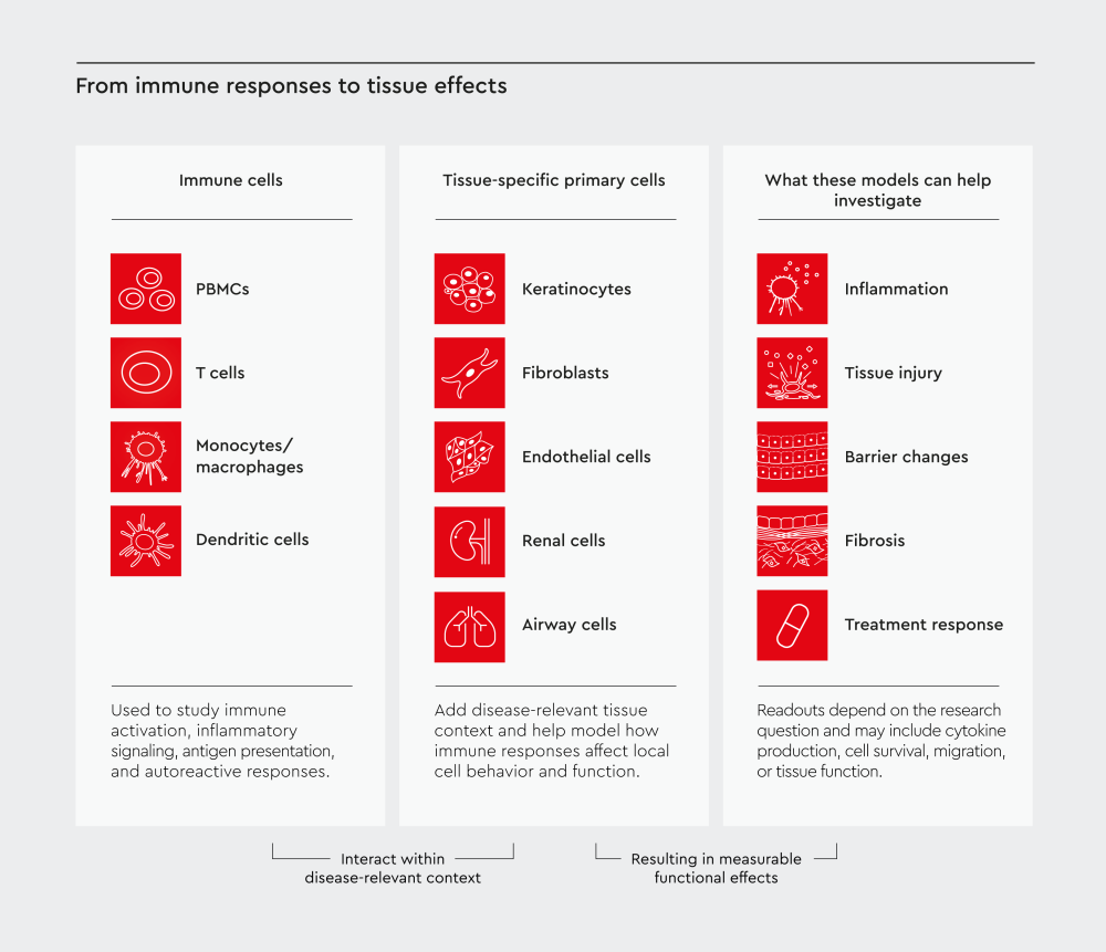
Figure 1. From immune responses to tissue effects.
Human primary-cell models in autoimmune research can combine immune cells with tissue-specific primary cells to investigate how immune responses affect local tissue behavior and function. Depending on the research question, these models can support studies of inflammation, tissue injury, barrier changes, fibrosis, and treatment response.
How immune cells capture different aspects of autoimmune responses
Different immune cell populations can provide insight into different aspects of autoimmune biology3,8:
- PBMCs: Broader studies of peripheral immune-cell responses.
- T cells: T-cell activation and antigen-specific activation or autoreactive responses.
- Monocytes and macrophages: Innate immune activity and inflammatory signaling.
- Dendritic cells: Antigen presentation and immune-cell activation.3,9
Typical readouts include cytokine and chemokine production, activation marker expression, proliferation, and differentiation.
How tissue-specific primary cells add the local disease context
In autoimmune disease, non-immune cells in the affected tissue can participate in or respond to ongoing inflammation, and this can influence the trajectory of the disease.10 Depending on the disease and research question, relevant tissue-specific cells may include keratinocytes, fibroblasts, endothelial cells, renal cells, pulmonary cells, and other cell types from the affected organ.
Recent research in rheumatoid and other autoimmune arthritic diseases highlights why this tissue context matters. A 2026 study identified distinct regulatory T-cell states enriched in inflamed synovial tissue rather than blood and showed that local macrophages and fibroblasts helped shape their functional state.11
Pairing immune and tissue-specific primary cells can therefore help you distinguish systemic immune-cell behavior from responses shaped by the local tissue environment.
Why combine immune cells with tissue-specific primary cells?
Combining immune cells with tissue-specific primary cells allows you to study the bidirectional crosstalk between immune responses and affected tissues. Immune cells can alter tissue-cell behavior, while tissue cells can amplify or regulate inflammatory responses.10
Depending on the research question, these interactions can be investigated using:
- direct co-culture when cell-cell contact contributes to the response
- indirect co-culture when reciprocal soluble signaling is relevant while the cell populations remain physically separated
- conditioned-medium experiments to investigate whether soluble factors released by one cell population are sufficient to affect another
- cytokine-stimulated tissue cells to study responses to defined inflammatory signals
- migration assays to investigate immune-cell recruitment
- cytotoxicity assays to assess immune-mediated cell injury
Recent rheumatoid arthritis research illustrates the value of this approach. In a 2026 study, researchers co-cultured human PBMCs with fibroblast-like synoviocytes from rheumatoid arthritis patients to investigate immune cell responses in a disease-relevant synovial environment.12
Not every autoimmune study needs a multicellular model. If immune–tissue crosstalk is central to your research question, however, combining relevant human cell types can reveal inflammatory and tissue responses that a single-cell-type model cannot directly capture.
Can primary cells be used for autoimmune drug screening?
Human primary cells can also support autoimmune drug screening and functional assays by showing how candidate treatments affect immune and tissue-cell responses. Relevant readouts may include13,14:
- inflammatory signaling and cytokine production
- immune-cell activation
- cytotoxicity or tissue-cell survival
- barrier or tissue function
- fibrotic responses
The most appropriate readout should reflect the mechanism of action and the biological response the treatment is intended to modify.15,16
How should donor variability be handled in autoimmune research?
Donor variability is both a challenge to experimental standardization and a source of biologically relevant information in human primary-cell research.17,18
The key is to decide whether your study should minimize variability or capture it.
When designing a study, consider:
- Number of donors: Use independent donors when the aim is to assess inter-individual variability. Replicates from the same donor do not capture donor-to-donor variability.
- Donor type: Healthy donor cells can provide a defined baseline for studying experimentally induced immune or inflammatory responses, while disease-relevant donor cells may be more appropriate when the aim is to investigate disease-associated phenotypes or patient heterogeneity.
- Donor characteristics: Consider factors such as age, sex, disease status, or other relevant characteristics where these could influence the response.
- Matched cells: Where interactions between different cell types are being studied, cells from the same donor may help control for differences in genetic background. Intentionally mismatched cells may instead be appropriate when allogeneic or HLA-dependent interactions are part of the research question.
Rather than treating donor variability only as experimental noise, you can incorporate it into the study design when human biological diversity is relevant to the question.17,18
When does HLA type matter in autoimmune research?
Human leukocyte antigen (HLA) type becomes particularly important when antigen presentation or HLA-restricted immune responses are part of the mechanism being studied.17,19
This can include:
- autoreactive T-cell responses
- disease-associated HLA alleles
- antigen-presentation assays
- HLA-restricted mechanisms or targets
- donor stratification
- matched or intentionally mismatched immune-cell systems
Not every autoimmune model requires HLA-typed cells. For general inflammatory or functional assays that do not depend on HLA-mediated interactions, HLA typing may not be a defining experimental variable.
For studies where HLA context is important, HLA-typed human primary cells can provide donor-defined material for investigating immune interactions and response patterns. See also our article on HLA in research for more insights on this topic.
What makes an autoimmune model physiologically relevant?
Physiological relevance does not necessarily mean greater model complexity. A useful autoimmune model includes the human cellular and biological context required to represent the mechanism being studied.
Before setting up an autoimmune model, consider the following:
- Biological mechanism: What immune or tissue response needs to be represented?
- Cellular context: Which immune cells and, where relevant, tissue-specific cells are involved?
- Interaction format: Does the mechanism require direct cell contact, soluble signaling, migration, or a defined inflammatory stimulus?
- Donor context: Could donor characteristics or donor-to-donor variability influence the response?
- HLA requirements: Is HLA relevant to the mechanism being studied?
- Functional readout: Which measurable response will show whether the biological effect has occurred?
The table below summarizes how you can use different cellular models to answer different autoimmune research questions3:
| Research focus | Possible cellular components | Donor / HLA considerations | Example readouts |
|---|---|---|---|
| Immune activation | Relevant primary immune cells | Multiple donors may help assess inter-individual responses | Cytokines, activation markers, proliferation |
| Tissue response to inflammation | Tissue-specific primary cells | Donor characteristics may influence tissue responses | Inflammatory signaling, tissue function, cell injury |
| Immune–tissue interactions | Immune + tissue-specific cells | Matched donor cells may be useful where relevant | Crosstalk, migration, tissue injury or protection |
| Antigen-specific responses | Immune and/or antigen-presenting cells | HLA may be an important experimental variable | Antigen presentation, T-cell activation, cytokine responses |
| Treatment response | Model depends on the therapeutic mechanism | Multiple donors may reveal response variability | Changes in inflammation, cytotoxicity or tissue function |
Table 1. Examples of cellular components and experimental considerations for different autoimmune research questions.
The goal is not to build the most elaborate model possible, but to include the cellular and donor context needed to answer your specific question.
Bringing your autoimmune model together
Ultimately, autoimmune model design is about matching the biological context to the research question. Once you have defined the mechanism, relevant cell types, donor context, and functional readout, the next step is to identify cells and culture conditions that match those requirements.
Explore our blood and immune cell portfolio and immunology research area to find human cell types and donor options that fit your experiments.
Looking for the right cells for your autoimmune model? Whether you're studying immune activation, tissue inflammation, or immune-tissue crosstalk, selecting the right human primary cells is critical. Explore our portfolio of blood, immune, and tissue-specific primary cells to build physiologically relevant autoimmune disease models.
Frequently asked questions
References
Expand
- Ahluwalia A, Fletemeyer B, Chirico G, et al. What’s in a NAM? Lab Anim. Published online 2026. doi:10.1038/s41684-026-01731-8
- Segeritz CP, Vallier L. Cell culture. In: Basic Science Methods for Clinical Researchers. Elsevier; 2017:151-172. doi:10.1016/B978-0-12-803077-6.00009-6
- Hammel JH, Zatorski JM, Cook SR, Pompano RR, Munson JM. Engineering in vitro immune-competent tissue models for testing and evaluation of therapeutics. Adv Drug Deliv Rev. 2022;182:114111. doi:10.1016/j.addr.2022.114111
- Ejma-Multański A, Wajda A, Paradowska-Gorycka and treatment of autoimmune connective tissue diseases. Cells. 2023;12(20):2489. doi:10.3390/cells12202489
- Zhu L, Sun W, Chen K, et al. Single-cell landscape of immune cells in multiple autoimmune diseases. iScience. 2026;29(1):114515. doi:10.1016/j.isci.2025.114515
- Wu X, Wu MA, Zou J, Kleinstreuer N, Wu JC. Reimagining human-centric drug development with new approach methodologies. Science. 2026;392(6796):371-378. doi:10.1126/science.aeb0045
- Şen B, Balcı-Peynircioğlu B. Cellular models in autoinflammatory disease research. Clin Transl Immunol. 2024;13(1):e1481. doi:10.1002/cti2.1481
- Song X, Liang H, Nan F, et al. Autoimmune diseases: molecular pathogenesis and therapeutic targets. MedComm. 2025;6(7):e70262. doi:10.1002/mco2.70262
- Patel AA, Ginhoux F, Yona S. Monocytes, macrophages, dendritic cells and neutrophils: an update on lifespan kinetics in health and disease. Immunology. 2021;163(3):250-261. doi:10.1111/imm.13320
- Sisto M, Lisi S. Immune and non-immune inflammatory cells involved in autoimmune fibrosis: new discoveries. J Clin Med. 2023;12(11):3801. doi:10.3390/jcm12113801
- Koh B, Gal Oz ST, Sato R, et al. Functional and dysfunctional T regulatory cell states in human tissues in RA and other autoimmune arthritic diseases. Nat Immunol. 2026;27(7):1449-1461. doi:10.1038/s41590-026-02540-4
- Bruci D, Lowin T, Fritz G, Pongratz G. Sublethal DNA damage switches off B cell effector programs in an RA-FLS-PBMC co-culture. Cell Death Discov. 2026;12(1):161. doi:10.1038/s41420-026-03021-1
- Albert-Vega C, Tawfik DM, Trouillet-Assant S, Vachot L, Mallet F, Textoris J. Immune functional assays, from custom to standardized tests for precision medicine. Front Immunol. 2018;9:2367. doi:10.3389/fimmu.2018.02367
- Yang Y, Liu J, Liu J, et al. Advances in immune cell-based therapeutic agents for the treatment of inflammation-related diseases. Acta Pharm Sin B. 2026;16(5):2794-2837. doi:10.1016/j.apsb.2026.01.013
- Yi S, Wu C, McIntosh A, Santaella ME, Robinson TM, Liao MZ. Cell and gene therapy: transforming treatment paradigms for patient-centric care. Clin Transl Sci. 2025;18(12):e70430. doi:10.1111/cts.70430
- Solimani F, Amagai M, Bollard CM, Payne AS. Clinical progress of engineered cellular immunotherapies for autoimmunity. Nat Biotechnol. 2026;44(4):547-562. doi:10.1038/s41587-026-03001-x
- Gibson D, Leonforte C, Madrigal A. Strategies for dealing with donor variability. Cell Gene Ther Insights. Published online 2018. doi:10.18609/cgti.2018.087
- Hoang Nguyen KH, Le NV, Nguyen PH, Nguyen HHT, Hoang DM, Huynh CD. Human immune system: Exploring diversity across individuals and populations. Heliyon. 2025;11(2):e41836. doi:10.1016/j.heliyon.2025.e41836
- Bodis G, Toth V, Schwarting A. Role of human leukocyte antigens (HLA) in autoimmune diseases. Rheumatol Ther. 2018;5(1):5-20. doi:10.1007/s40744-018-0100-z
Related resources