Air-liquid interface culture (ALI) allow scientists to generate stable and functional in vitro 3D human airway cell models that closely mimic respiratory tract epithelia. With such models, researchers can investigate physiological and pathological processes of the respiratory tract. They can study interactions between epithelial cells and respiratory pathogens or air pollutants, and observe how cells respond to aerosolized drugs.
We do not think about it. We almost do not notice it, but every minute we inhale and exhale seven to eight liters of air. This air contains the oxygen we need to live. But it also can introduce dust, pollutants and potentially harmful pathogens into our bodies. It is the job of the epithelial cells in our airways to build a barrier and make sure that those agents are quickly removed so as to avoid damage to our lungs. However, pulmonary diseases such as asthma or chronic obstructive pulmonary diseases (COPD) impair this “cleaning” ability of the airway epithelium, and the consequences can be very severe. In order to get a closer look into what is happening inside our airways, researchers need to replicate the native environment in vitro as closely as possible. With the current strict regulations and the emphasis on the so-called 3R principle aimed at replace, reduce and refine the use of animal testing, there is a growing need for physiologically relevant models of the respiratory epithelium. Air-liquid interface (ALI) systems closely mimic the in vivo characteristics of the pulmonary epithelial cells and therefore represent an optimal alternative to animal studies. Overall, ALI systems can provide valuable information on physiological and pathological processes in the respiratory tract, with their potential applications ranging from the study of pulmonary diseases to the fight against infectious agents to the investigations of the effects of air pollution on the respiratory system. Yet, as with every in vitro system, there are several challenges scientists need to overcome to ensure that the ALI system used is validated, standardized and offers relevant and reproducible results.A major challenge: replicating the complex anatomy of human lungs in vitro
With approximately 80 m², the surface of our lungs is the size of a tennis court and is paved with not just one but with multiple cell types. There are goblet cells, basal cells and ciliated epithelial cells, among others (Miller and Spence, 2017). The reason for this variety of cell types is the multiple tasks the airway epithelium has to accomplish. First, every hour it has to deal with up to 25 million inhaled particles and prevent them from entering and damaging the body. Second, goblet cells and sero-mucous glands produce mucus, a very effective defensive tool. This viscoelastic gel forms a protective coating which captures inhaled microorganisms. Then a mechanism called mucociliary clearance transports any foreign particles to the mouth to clear them away (Rogers, 2007). Last, epithelial cells produce several cytokines that play a role in innate and adaptive immune response mechanisms (Mertens et al., 2017). Because epithelial cells perform so many tasks, reestablishing a well-functioning respiratory epithelium in vitro is challenging. Two-dimensional cell cultures present several disadvantages, such as the loss of tissue-specific architecture, the absence of cellular differentiation and damage of barrier integrity. All of these features are very important when investigating respiratory diseases. In fact, the ability to undergo differentiation, develop cilia and produce mucus are key features of human bronchial epithelial cells. Often these features are lost in 2D in vitro cultures, where cells show fast dedifferentiation, high senescence and low proliferation (Ramirez et al., 2004). Moreover, monolayers do not exhibit the typical pseudostratified morphology and do not form tight junctions, so they are more sensitive to disruptions. The use of airway derived cell-lines also presents some limitations as those are often cancer-derived, and their physiological processes do not always resemble the in vivo characteristics of normal epithelial cells (Ryner et al, 2019). Due to these reasons, 3D primary human airway epithelial cultures have gained more and more interest recently and are increasingly used for in vitro scientific studies. ALI culture systems manage to overcome many of the challenges of 2D cultures and have many advantages when compared to submerged culture systems. ALI cultures exhibit a pseudostratified morphology, undergo mucociliary differentiation, secrete mucin and form tight junctions. Also, the extracellular environment is similar to the in vivo conditions (Lacroix et al., 2018).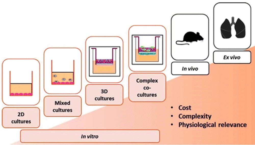
ALI cultures: powerful models of the human airways
Cell cultures performed at the air-liquid interface facilitate the establishment of stable and functional 3D models of the respiratory tract. In these models, the basal side of the cells is in contact with the culture medium, and the apical side is in contact with the air. Therefore, the created in vitro scenario is close to what occurs in vivo (Lacroix et al., 2018). To establish ALI cultures, primary epithelial cells are first seeded on plastic culture vessels, where they can expand until they reach 70-80% confluence. After this first 2D expansion phase, cells are transferred to compartmentalized culture systems on porous membranes. Here, cells are submerged in culture medium and start proliferating in 3D until reaching confluence. Finally, they are ‘air-lifted,’ i.e. exposed to the surrounding air, and nutritive supply is provided only at the basolateral cell pole. This system allows morphological and functional cell differentiation and the formation of a pseudostratified epithelium with full basoapical polarity. The reconstituted epithelium shows many features observed in vivo – such as the production of cilia and mucus (Chen and Schoen, 2019). ALI systems also support the development of tight junctions which are critical for the epithelial barrier function. The measurement of the transepithelial electrical resistance (TEER) allows the assessment of the barrier integrity. The measurement of TEER is an important strategy to confirm epithelial polarization and therefore the maintenance of normal function of airway epithelium. It can be used routinely without damaging the cells and allows the study of the same cells in different setups (Papazian et al., 2016). For this purpose, an alternating current is applied to the cell layer, and the electrical resistance is then calculated (Knowles et al., 1982). Due to their physiological relevance, ALI systems are widely used and are now an essential tool in respiratory research. However, the lack of validation and standardization in the field hinders the comparison of different studies. In fact, the use of heterogeneous cultivation protocols, cells, scaffolds and media by different research groups complicates the inter-laboratory reproducibility of the results (Zscheppang et al., 2017).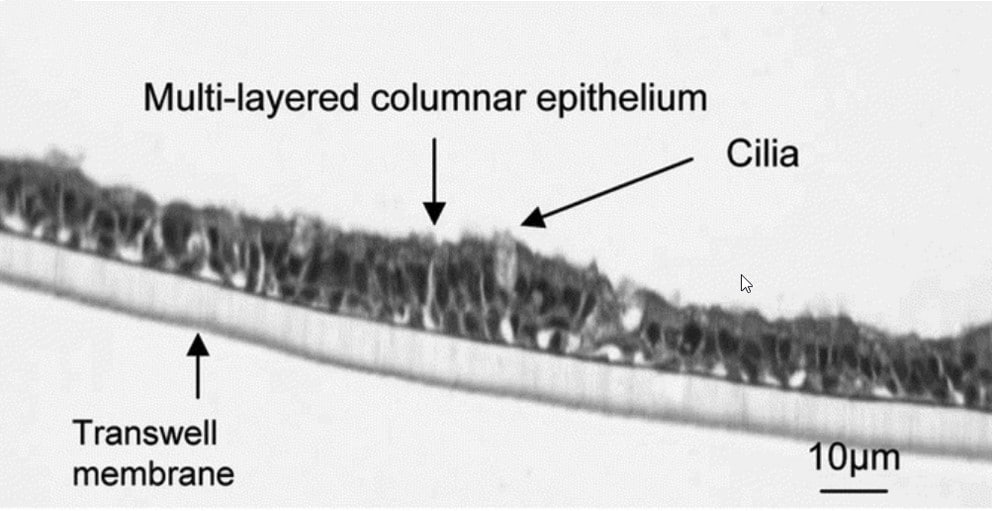
Standardization: a key requirement for creating relevant culture systems
Standardization and validation of methods based on original human material become even more important when investigation results have clinical relevance. As animal models do not exactly mirror human physiology, standardized experimental approaches in human models significantly contribute to expediting translational processes while meeting, at the same time, ethical concerns regarding the usage of animal testing. Robust procedures are also a prerequisite for acceptance by regulatory authorities and pharmaceutical industries (Zscheppang et al., 2017). Using defined culture media and pre-screened cells is a concrete step forward towards the establishment of such standardized cultures. The use of serum and bovine pituitary extract (BPE)-free culture media allows the generation of predictive and reproducible 3D airway model systems and offers several advantages, such as:- Defined and controlled culture conditions
- Reduced variability in qualitative and quantitative culture medium composition
- Reduced risks of microbial contamination
- Higher resemblance of in vivo physiological conditions
- Reduction or even avoidance of the suffering of fetuses and animals
Using airway epithelial cell systems to better understand pulmonary diseases
Pulmonary diseases such as chronic obstructive pulmonary disease (COPD), asthma, pneumonia and lung cancer have a huge global impact, affecting millions of people worldwide. As well, billions of people are exposed to indoor toxic smoke, outdoor pollutant air and tobacco smoke. These data make it clear why there is a high need for establishing reliable in vitro research models to study both pathological processes in the airway epithelium and the influence of infectious agents and toxic substances on epithelial cells. Further, dysfunctions of epithelial cells are often the cause of chronic lung diseases such as asthma and chronic obstructive pulmonary disease (COPD). ALI systems use primary human cells to model such diseases and allow a better understanding of the underlying pathological changes. Moreover, cultured cells can be exposed to pathogens, aerosolized medications and toxic substances, and air pollution can be used to model phases of acute exacerbation or observe the effects of therapeutic drugs (Mertens et al., 2017). In fact, when studying the effects of drugs used in inhalation therapy, ALI systems offer a more physiological model than submerged cultures. When drugs are dissolved in the medium, they cover the whole cell and do not mimic the in vivo scenario where drugs, e.g. applied as an aerosol, only come in contact with the air-facing side of the epithelial cells (Lenz et al., 2014). ALI cell culture models can also be used to assess the effect of infectious pathogens such as viruses and bacteria on airway epithelial cells. As animal models are not always natural hosts of human pathogens, it is crucial to develop standardized systems which closely resemble human airways. During the current SARS-CoV-2 pandemic, for example, researchers have used ALI culture systems to study host responses to the infections and to screen therapeutic drugs. In a recent study, Mulay et al. isolated human epithelial cells from trachea and upper bronchi and differentiated them at ALI for 16–20 days until a pseudostratified mucociliary epithelium was formed. The team then infected these ALI cultures with SARS-CoV-2 and observed virus replication and gene expression in infected cells. The proximal airway ALI cultures, together with 3D alveolar organoids, were also used to study the effect of a selected panel of drugs such as IFNB1, Remdesivir and Hydroxychloroquine on viral infection and replication. Toxicology research also greatly benefits from the use of ALI exposure systems. Scientists can investigate cellular reactions and cell-cell interactions following exposure to toxic substances, air pollutants or drugs. Moreover, they can use these systems to better understand how aerosolized or gaseous substances contribute to the development of lung diseases (Upadhyay et al., 2018). The advantage of ALI systems is that they provide a method to challenge your cultured epithelium with any agent of interest. Either air pollutants, toxic substances, viruses, bacteria or drugs can be used to observe the reactions of the cells to these agents. The system is a valid alternative to in vivo experiments or classic in vitro methods. Further development of ALI-models is aimed at bringing the systems even closer to human physiology, increasing standardization and establishing validated methods. The use of ALI systems for 3D organoid culture could also significantly improve human relevance of the results obtained in in vitro studies and provide important insights into the dynamic processes in our airways. Such powerful tools will allow researchers to obtain more detailed insights in the highly specialized barrier mechanisms of the lung epithelia.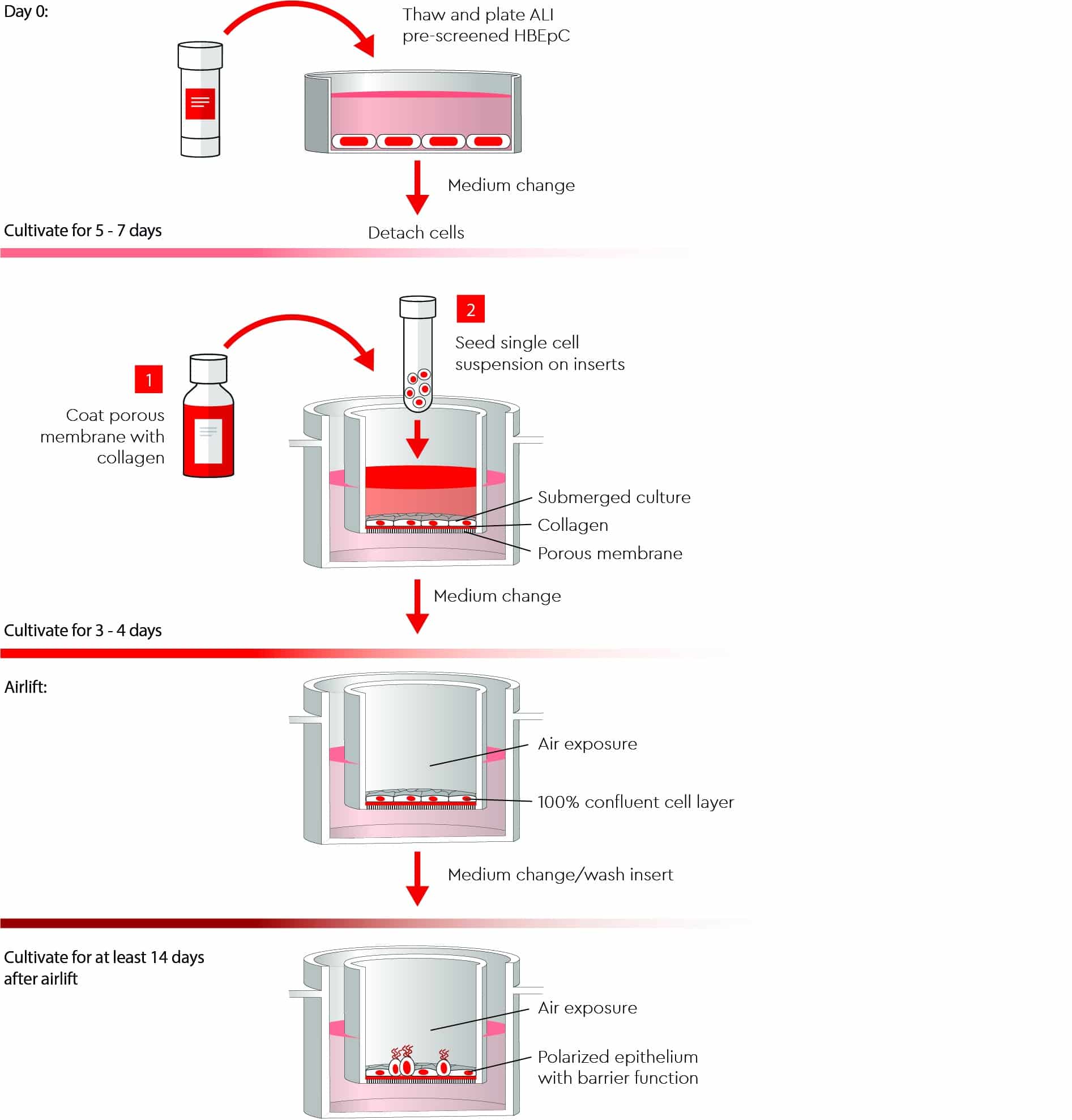
Frequently asked questions about ALI
What are ALI pre-screened cells?
The pre-screening of primary human bronchial epithelial cells for ALI cultures guarantees an optimal barrier function and minimizes lot-to-lot variations. Cells are collected from surgical samples after obtaining the consent of the patients. Patients are aware that their consent to donate cells contributes to the reduction of animal experiments. Screening is then performed by analyzing the transepithelial electrical resistance (TEER) of the cells over a time period of at least 14 days. TEER values of over 500 Ω*cm² are accepted. Moreover, a strict quality control process ensures that the cells meet high quality standards. PromoCell human bronchial epithelial cells are low in passages and have very good proliferation and barrier forming function capacities, with a minimum of 15 population doubling.What are the advantages of using a standardized ALI culture medium?
Standardized media display less lot-to-lot variations and help to maximize your specific test reproducibility. Moreover, BPE- and serum-free media provide more test-to-test consistency and eliminate possible contaminations. The PromoCell ALI-airway medium is a valuable tool for basic respiratory research, toxicity studies, infectious disease research and drug development. It is easy to use, optimized for 3D cell cultures and allows the formation of tight junctions and, therefore, a long-lasting barrier function.When do you recommend starting ‘air-lifting’ the cells?
We recommend a seeding density of 150.000 living cells/cm2 on the Transwell inserts in Airway Epithelial Cell Growth Medium. Twenty-four hours after seeding, a medium change has to be performed. At this point you should check the cell layer under a microscope, which should be nearly confluent. After three more days of culture, the cell layer should form a monolayer with nearly 100% confluence. When you are sure that the cell layer is confluent, you can change the medium in the basal chamber and keep the apical chamber empty to airlift the cells and expose them to the air.Discover how PromoCell can support you in establishing predictive and reproducible air liquid interface culture systems
PromoCell has introduced a new ALI Medium and pre-screened guaranteed primary human bronchial epithelial cells for ALI culture systems.
Explore our portfolio
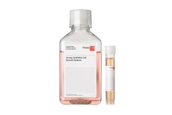
Serum-free cell culture medium for epithelial cells from large air passages.
Primary Human Bronchial Epithelial Cells isolated from the surface epithelium of human bronchi.
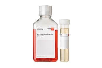
Serum-free and BPE-free cell culture medium for an optimal and standardized culture of human bronchial epithelial cells at the air-liquid interface (ALI).
Related products