

Citations
PromoCell uses the Bioz AI engine to display product citations. This content is currently blocked due to your cookie preferences.
Human Dermal Blood Endothelial Cells (HDBEC)
Human Dermal Blood Endothelial Cells (HDBEC) adult donor
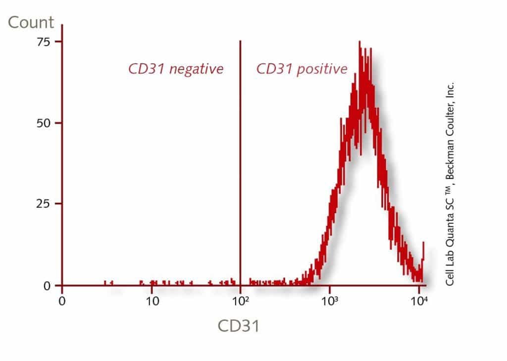
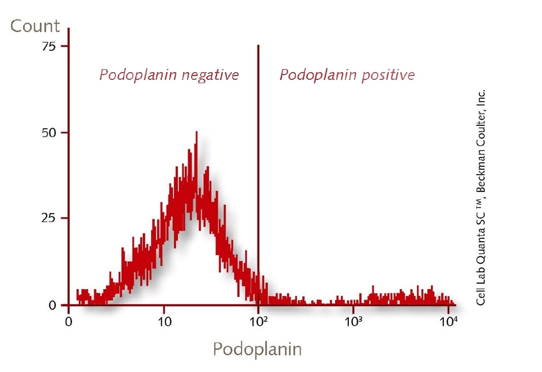
Primary Human Dermal Blood Endothelial Cells (HDBEC) are a subpopulation of the Human Dermal Endothelial Cells. They are isolated from the dermis of juvenile foreskin and adult skin (different locations) from a single donor. The cells are analyzed positive for CD31 and negative for podoplanin by flow cytometric analysis.
Human Dermal Lymphatic Endothelial Cells (HDLEC) from the same donor are available on request.
Blood Endothelial Cells have a key function in physiological processes like vessel tonus, capillary permeability, blood coagulation, fibrolysis, and angiogenesis. They are also useful for disease studies of atherosclerosis, tumor genesis, and thrombosis.
- Request our GMP grade cell culture media for endothelial cells.
- Our HDBEC are now also available from HLA-typed donors.
Recommended plating density | 10000 - 20000 cells per cm2 |
Passage after thawing | P2 |
Tested markers | Podoplanin positive, CD31 positive |
Guaranteed population doubling | > 15 |


We use a classification system similar but not identical to the Fitzpatrick Skin Classification. The Fitzpatrick classification has six different categories (phototypes I-VI) which correlate with the level of skin pigmentation (melanin) and sunburn following sun exposure. Fitzpatrick I corresponds with the lightest of skin complexions, while Fitzpatrick VI corresponds with the darkest skin.
- I: Pale white skin, blue/hazel eyes, blond/red hair, always burns, does not tan
- II: Fair skin, blue eyes, burns easily, tans poorly
- III: Darker white skin, tans after initial burn
- IV: Light brown skin, burns minimally, tans easily
- V: Brown skin, rarely burns, tans darkly easily
- VI: Dark brown or black skin, never burns, always tans darkly
At PromoCell, we have knowledge of the patients’ skin color (white, brown or black skin), color of eyes and hair, but we don't have any details about the burning/tanning abilities. We therefore classify our tissue donors as follows:
- Light (comprising phototypes I and II)
- Moderate (comprising phototypes III and IV)
- Dark (comprising phototypes V and VI)
Information on the phototype is available for most cell lots isolated from juvenile or adult skin.
Short protocol:
- Trypsinize the cells as usual
- Centrifuge and resuspend in suitable cold freezing medium at a density of 1-4 x 10⁶ cells/ml
- Cool down the cells slowly to -80°C (approx. -1°C per min). We recommend to use "Mr. Frosty" from Nalge or "CoolCell" from Biocision; which both provide gradual and controlled cooling rates when placed in a -80°C freezer overnight.
- Transfer the vials into liquid nitrogen for long-term storage
- Mesenchymal Stem Cells (C-12974/C-12971/C-12977) need Fibronectin-coating when grown in PromoCell MSC Growth Medium XF (C-28019) and when differentiated in MSC Neurogenic (C-28015); Adipogenic (C-28016); or Osteogenic (C-28013) Differentiation Media.
- Human monocyte-derived macrophages (C-12914/C-12916/C-12915/C-12917) must be seeded into Fibronectin-coated culture vessels in combination with PromoCell's M1- and M2-Generation Media XF (C-28055; C-28056).
- For efficient induction of osteoblast mineralization with PromoCell's Osteoblast Mineralization Medium (C-27020); the TC plates should be pre-coated with collagen type I.
- Liquid phase storage provides a consistent temperature of -196°C; a longer holding time and a greater vial capacity but involves the risk of contamination issues.
- Storage in the gas phase is very safe with respect to contaminations but the holding time of the cells is shorter and the vial capacity is reduced.
General protocol for recovery of anchorage-dependent primary cells: Remove vial from liquid nitrogen; transport it on dry ice to the cell culture lab. Thaw in a 37°C waterbath for approx. 2 min; until it is just defrosted. Keep the vial immersed in water until just below the screw cap during thawing and only remove it shortly after approx. 90 sec to check the progress. Do not repeatedly insert and remove the vial while thawing in water. Carefully disinfect the vial with plenty of 70% EtOH under the laminar flow hood and aseptically transfer the thawed suspension into an appropriate TC dish with growth medium (pre-warmed in the incubator for > 30 min). The cells usually attach within a few hours. Perform media change after 24 hrs at the latest to remove residual DMSO from the freezing media. Additional information can be found in the instruction manuals of our cells.
The study of angiogenesis has been significantly advanced by the ability to culture endothelial cells in vitro. Initially large vessel ECs such as those isolated from the human umbilical vein (HUVEC) were used for these studies but increasingly it has been recognized that microvascular endothelial cells are a more appropriate model since angiogenesis involves microvessels rather than large vessel ECs.


PromoCell uses the Bioz AI engine to display product citations. This content is currently blocked due to your cookie preferences.
Recommended products

Endothelial Cell Growth Medium MV
Low-serum cell culture medium for endothelial cells from microvascular vessels, the coronary artery, and the aorta.