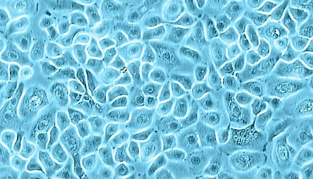
Human Tracheal Epithelial Cells (HTEpC)
 Powered by Bioz
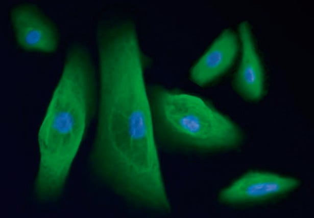
Primary Human Tracheal Epithelial Cells (HTEpC) are isolated from the surface epithelium of human trachea and stain positive for cytokeratin. The respiratory epithelia are responsible for the lubrication of the lungs, the maintenance of humidity, and the cleaning of the respiratory tract. They are an important target for drugs, toxins, and carcinogens. In addition, they are involved in many diseases such as cystic fibrosis. Consequently, HTEpC are useful for investigating the function and pathology of the respiratory system.
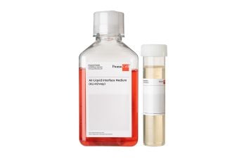
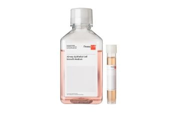
- Request our GMP grade cell culture media for airway epithelial cells.
- Our HTEpC are now also available from HLA-typed donors.
| Recommended plating density | 10,000 - 15,000 cells per cm2 |
| Passage after thawing | P2 |
| Tested markers | Cytokeratin positive |
| Guaranteed population doubling | > 15 |
| Recommended culture media* | C-21260, C-21050, C-21040 |
*The catalog numbers in this table are for media in ready-to-use packaging.
PromoCell uses the Bioz AI engine to display scientific references for this product. This content is currently blocked because functional cookies are disabled.
Interested in our scientific references?
Click on the following link to load the content or enable functional cookies in the consent settings.
By clicking "Load content now", you agree to load content from Bioz, a third-party provider. This will set cookies on your device without changing your saved cookie preferences.