


Technical library
Items 133-144 of 283 Results
 PromoCell_Best-Practices-in-Cell-Culture
tools-for-respiratory-research_brochure
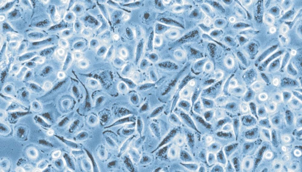
Standardized culture of assay-ready and fully functional human primary macrophages
Trypsin Neutralizing Solution - 30 ml
Trypsin Neutralizing Solution - 125 ml
Trypsin Neutralizing Solution - 250 ml
PromoCell_Best-Practices-in-Cell-Culture
tools-for-respiratory-research_brochure
Standardized culture of assay-ready and fully functional human primary macrophages
Trypsin Neutralizing Solution - 30 ml
Trypsin Neutralizing Solution - 125 ml
Trypsin Neutralizing Solution - 250 ml
A concentration > 10% FBS is needed to completely inactivate the trypsin. As most of PromoCell's growth media are serum-reduced or serum-free, the use of a trypsin inhibitor like TNS is highly recommended.
PromoCell_Best-Practices-in-Cell-Culture
tools-for-respiratory-research_brochure
Standardized culture of assay-ready and fully functional human primary macrophages
Endothelial Cell Growth Medium (Ready-to-use)
Endothelial Cell Growth Medium 2 (Ready-to-use)
Human Pulmonary Artery Endothelial Cells (HPAEC)
Human Saphenous Vein Endothelial Cells (HSaVEC)
Human Umbilical Artery Endothelial Cells (HUAEC)
Human Umbilical Vein Endothelial Cells (HUVEC) pre-screened
Human Umbilical Vein Endothelial Cells (HUVEC) pooled
Human Umbilical Vein Endothelial Cells (HUVEC) single donor
The standard medium for isolation and propagation of our HUVEC, HUAEC, HPAEC, and HSaVEC is Endothelial Cell Growth Medium (C-22010). It contains ECGS, an extract from bovine hypothalamus which has mitogenic effects on endothelial cell proliferation. Scientists who prefer a more defined Growth Medium can use Endothelial Cell Growth Medium 2 (C-22011). In this medium, ECGS is replaced by VEGF, IGF, and additional bFGF and EGF to stimulate endothelial cell growth.
PromoCell_Best-Practices-in-Cell-Culture
tools-for-respiratory-research_brochure
Standardized culture of assay-ready and fully functional human primary macrophages
Endothelial Cell Growth Medium MV2 (Ready-to-use)
PromoCell's HDLEC are isolated from skin (dermis). When isolated from juvenile donors (C-12216), the exact localization is foreskin. When derived from adult donors (C-12217), the localization depends on the type of surgery, e.g. breast or temple. You can find the information on the exact localization in the Certificate of Analysis.
Our HDLEC are tested to be positive for CD31, podoplanin, and ac-LDL uptake and are delivered in P2. The recommended culture medium is Endothelial Cell Growth Medium MV2 (C-22022).
PromoCell_Best-Practices-in-Cell-Culture
tools-for-respiratory-research_brochure
Standardized culture of assay-ready and fully functional human primary macrophages
Human Umbilical Vein Endothelial Cells (HUVEC) pooled
Endothelial Cell Growth Medium (Ready-to-use)
PromoCell's Endothelial Cell Growth Medium (C-22010) is a complete medium that can be used for the culture of HUVEC after addition of the SupplementMix. Addition of extra FCS is not necessary. The SupplementMix contains FCS (2% v/v final concentration), recombinant growth factors, hormones, and a bovine brain extract that together have mitogenic effects on endothelial cells.
PromoCell_Best-Practices-in-Cell-Culture
tools-for-respiratory-research_brochure
Standardized culture of assay-ready and fully functional human primary macrophages
Human Umbilical Vein Endothelial Cells (HUVEC) single donor
Human Umbilical Vein Endothelial Cells (HUVEC) pooled
Our HUVEC single donor (C-12200) are isolated from a single umbilical cord, propagated in primary culture, and frozen down at subconfluency. For the preparation of HUVEC-pooled (C12203), we simultaneously isolate the cells from 2-4 umbilical cords and grow them in separate tissue culture dishes. The cells are pooled after trypsinization given that their growth rates are comparable. After thawing, our HUVECs (single donor and pooled) are both in P1. The recommended media are Endothelial Cell Growth Medium (C-22010) or Endothelial Cell Growth Medium 2 (C-22011). With respect to cell growth, HUVEC-pooled tend to have a more heterogeneous morphology with slightly more elongated cells but the doubling times are comparabel for both types (typically 18-36 hrs per doubling).
PromoCell_Best-Practices-in-Cell-Culture
tools-for-respiratory-research_brochure
Standardized culture of assay-ready and fully functional human primary macrophages
Endothelial Cell Growth Medium SupplementMix
Endothelial Cell Growth Medium SupplementPack
Endothelial Cell Growth Medium Kit
Endothelial Cell Growth Medium (Ready-to-use)
Endothelial Cell Growth Medium 2 SupplementMix
Endothelial Cell Growth Medium 2 SupplementPack
Endothelial Cell Growth Medium 2 Kit
Endothelial Cell Growth Medium 2 (Ready-to-use)
Endothelial Cell Growth Medium MV SupplementMix
Endothelial Cell Growth Medium MV SupplementPack
Endothelial Cell Growth Medium MV Kit
Endothelial Cell Growth Medium MV (Ready-to-use)
The source of our heparin is ex porcine mucosa.
PromoCell_Best-Practices-in-Cell-Culture
tools-for-respiratory-research_brochure
Standardized culture of assay-ready and fully functional human primary macrophages
Adipocyte Nutrition Medium (Ready-to-use)
Adipocyte Basal Medium
Airway Epithelial Cell Growth Medium (Ready-to-use)
Chondrocyte Basal Medium
Chondrocyte Growth Medium (Ready-to-use)
Endothelial Cell Growth Medium (Ready-to-use)
Endothelial Cell Growth Medium 2 (Ready-to-use)
Endothelial Cell Growth Medium MV (Ready-to-use)
Endothelial Cell Growth Medium MV2 (Ready-to-use)
Fibroblast Growth Medium (Ready-to-use)
Fibroblast Growth Medium 2 (Ready-to-use)
Fibroblast Growth Medium 3 (Ready-to-use)
Follicle Dermal Papilla Cell Growth Medium (Ready-to-use)
Human Aortic Adventitial Fibroblasts (HAoAF)
Human Aortic Endothelial Cells (HAoEC)
Human Aortic Smooth Muscle Cells (HAoSMC)
Human Bronchial Epithelial Cells (HBEpC)
Human Bronchial Smooth Muscle Cells (HBSMC)
Human Cardiac Fibroblasts (HCF)
Human Cardiac Microvascular Endothelial Cells (HCMEC)
Human Cardiac Myocytes (HCM)
Human CD14+ Monocytes (hMoCD14+-PB)
Human Chondrocytes (HCH)
Human Coronary Artery Endothelial Cells (HCAEC)
Human Dermal Blood Endothelial Cells (HDBEC) adult donor
Human Dermal Blood Endothelial Cells (HDBEC) juvenile foreskin
Human Dermal Lymphatic Endothelial Cells (HDLEC) adult donor
Human Dermal Lymphatic Endothelial Cells (HDLEC) juvenile foreskin
Human Dermal Microvascular Endothelial Cells (HDMEC) pre-screened
Human Dermal Microvascular Endothelial Cells (HDMEC) adult donor
Human Dermal Microvascular Endothelial Cells (HDMEC) juvenile foreskin
Human Follicle Dermal Papilla Cells (HFDPC)
Human M1 Macrophages (GM-CSF), Monocyte-derived, single donor, 1,5 Mio cells
Human M2 Macrophages (M-CSF), Monocyte-derived, single donor, 1,5 Mio cells
Human Mesenchymal Stem Cells from Adipose Tissue (hMSC-AT)
Human Mesenchymal Stem Cells from Bone Marrow (hMSC-BM)
Human Mesenchymal Stem Cells from Umbilical Cord Matrix (hMSC-UC)
Human Mononuclear Cells from Peripheral Blood (hMNC-PB), single donor, ultra-pure
Human Mononuclear Cells from Cord Blood (hMNC-CB), single donor, ultra-pure
Human Nasal Epithelial Cells (HNEpC)
Human Osteoblasts (HOB)
Human Pulmonary Artery Endothelial Cells (HPAEC)
Human Pulmonary Artery Smooth Muscle Cells (HPASMC)
Human Pulmonary Fibroblasts (HPF)
Human Pulmonary Microvascular Endothelial Cells (HPMEC)
Human Renal Cortical Epithelial Cells (HRCEpC)
Human Renal Epithelial Cells (HREpC)
Human Saphenous Vein Endothelial Cells (HSaVEC)
Human Skeletal Muscle Cells (SkMC)
Normal Human Epidermal Keratinocytes (NHEK) adult, pooled
Normal Human Epidermal Keratinocytes (NHEK) juvenile foreskin, pooled
Normal Human Epidermal Keratinocytes (NHEK) adult, single donor
Normal Human Epidermal Keratinocytes (NHEK) juvenile foreskin, single donor
Human Small Airway Epithelial Cells (HSAEpC)
Human Tracheal Epithelial Cells (HTEpC)
Human Tracheal Smooth Muscle Cells (HTSMC)
Human Umbilical Artery Endothelial Cells (HUAEC)
Human Umbilical Artery Smooth Muscle Cells (HUASMC)
Human Umbilical Vein Endothelial Cells (HUVEC) pre-screened
Human Umbilical Vein Endothelial Cells (HUVEC) pooled
Human Umbilical Vein Endothelial Cells (HUVEC) single donor
Human Umbilical Vein Endothelial Cells (HUVEC) isolated in Growth Medium 2, pooled
Human Uterine Fibroblasts (HUF)
Human Uterine Microvascular Endothelial Cells (HUtMEC)
Human Uterine Smooth Muscle Cells (HUtSMC)
Human White Preadipocytes (HWP) subcutaneous
Human White Preadipocytes (HWP) visceral
Keratinocyte Growth Medium 2 (Ready-to-use)
Keratinocyte Growth Medium 3
Mammary Epithelial Cell Growth Medium (Ready-to-use)
Melanocyte Growth Medium (Ready-to-use)
Myocyte Growth Medium (Ready-to-use)
Normal Human Dermal Fibroblasts (NHDF) adult donor
Normal Human Dermal Fibroblasts (NHDF) juvenile foreskin
Normal Human Epidermal Keratinocytes (NHEK) adult, isolated in Growth Medium 3, pooled
Normal Human Epidermal Keratinocytes (NHEK) juvenile foreskin, isolated in Growth Medium 3, pooled
Normal Human Epidermal Keratinocytes (NHEK) adult, isolated in Growth Medium 3, single donor
Normal Human Epidermal Keratinocytes (NHEK), juvenile foreskin isolated in Growth Medium 3, single donor
Normal Human Epidermal Melanocytes (NHEM)
Normal Human Epidermal Melanocytes (NHEM) juvenile foreskin, cultured in M3 Medium
Normal Human Epidermal Melanocytes (NHEM) adult donor, cultured in M3 Medium
Osteoblast Growth Medium (Ready-to-use)
Osteoblast Mineralization Medium
Preadipocyte Differentiation Medium Kit
Preadipocyte Growth Medium (Ready-to-use)
Renal Epithelial Cell Growth Medium 2 (Ready-to-use)
Skeletal Muscle Cell Growth Medium (Ready-to-use)
Skeletal Muscle Cell Differentiation Medium (Ready-to-use)
Small Airway Epithelial Cell Growth Medium (Ready-to-use)
Smooth Muscle Cell Growth Medium 2 (Ready-to-use)
Melanocyte Growth Medium M3 (Ready-to-use)
Generally, the medium should be changed every 2-3 days. Please note: Following thawing, the first medium change should be performed after 16-24 hours to prevent cell damage due to residual freezing medium.
PromoCell_Best-Practices-in-Cell-Culture
tools-for-respiratory-research_brochure
Standardized culture of assay-ready and fully functional human primary macrophages
Human Aortic Adventitial Fibroblasts (HAoAF)
Human Aortic Endothelial Cells (HAoEC)
Human Aortic Smooth Muscle Cells (HAoSMC)
Human Bronchial Epithelial Cells (HBEpC)
Human Bronchial Smooth Muscle Cells (HBSMC)
Human Cardiac Fibroblasts (HCF)
Human Cardiac Microvascular Endothelial Cells (HCMEC)
Human Cardiac Myocytes (HCM)
Human Chondrocytes (HCH)
Human Coronary Artery Endothelial Cells (HCAEC)
Human Dermal Blood Endothelial Cells (HDBEC) adult donor
Human Dermal Blood Endothelial Cells (HDBEC) juvenile foreskin
Human Dermal Lymphatic Endothelial Cells (HDLEC) adult donor
Human Dermal Lymphatic Endothelial Cells (HDLEC) juvenile foreskin
Human Dermal Microvascular Endothelial Cells (HDMEC) pre-screened
Human Dermal Microvascular Endothelial Cells (HDMEC) adult donor
Human Dermal Microvascular Endothelial Cells (HDMEC) juvenile foreskin
Human Follicle Dermal Papilla Cells (HFDPC)
Human Mesenchymal Stem Cells from Adipose Tissue (hMSC-AT)
Human Mesenchymal Stem Cells from Bone Marrow (hMSC-BM)
Human Mesenchymal Stem Cells from Umbilical Cord Matrix (hMSC-UC)
Human Nasal Epithelial Cells (HNEpC)
Human Osteoblasts (HOB)
Human Pericytes from Placenta (hPC-PL)
Human Pulmonary Artery Endothelial Cells (HPAEC)
Human Pulmonary Artery Smooth Muscle Cells (HPASMC)
Human Pulmonary Fibroblasts (HPF)
Human Pulmonary Microvascular Endothelial Cells (HPMEC)
Human Renal Cortical Epithelial Cells (HRCEpC)
Human Renal Epithelial Cells (HREpC)
Human Saphenous Vein Endothelial Cells (HSaVEC)
Human Skeletal Muscle Cells (SkMC)
Normal Human Epidermal Keratinocytes (NHEK) adult, pooled
Normal Human Epidermal Keratinocytes (NHEK) juvenile foreskin, pooled
Normal Human Epidermal Keratinocytes (NHEK) adult, single donor
Normal Human Epidermal Keratinocytes (NHEK) juvenile foreskin, single donor
Human Small Airway Epithelial Cells (HSAEpC)
Human Tracheal Epithelial Cells (HTEpC)
Human Tracheal Smooth Muscle Cells (HTSMC)
Human Umbilical Artery Endothelial Cells (HUAEC)
Human Umbilical Artery Smooth Muscle Cells (HUASMC)
Human Umbilical Vein Endothelial Cells (HUVEC) pre-screened
Human Umbilical Vein Endothelial Cells (HUVEC) pooled
Human Umbilical Vein Endothelial Cells (HUVEC) single donor
Human Umbilical Vein Endothelial Cells (HUVEC) isolated in Growth Medium 2, pooled
Human Uterine Fibroblasts (HUF)
Human Uterine Microvascular Endothelial Cells (HUtMEC)
Human Uterine Smooth Muscle Cells (HUtSMC)
Human White Preadipocytes (HWP) subcutaneous
Human White Preadipocytes (HWP) visceral
Normal Human Dermal Fibroblasts (NHDF) adult donor
Normal Human Dermal Fibroblasts (NHDF) juvenile foreskin
Normal Human Epidermal Keratinocytes (NHEK) adult, isolated in Growth Medium 3, pooled
Normal Human Epidermal Keratinocytes (NHEK) juvenile foreskin, isolated in Growth Medium 3, pooled
Normal Human Epidermal Keratinocytes (NHEK) adult, isolated in Growth Medium 3, single donor
Normal Human Epidermal Keratinocytes (NHEK), juvenile foreskin isolated in Growth Medium 3, single donor
Normal Human Epidermal Melanocytes (NHEM)
Normal Human Epidermal Melanocytes (NHEM) juvenile foreskin, cultured in M3 Medium
Normal Human Epidermal Melanocytes (NHEM) adult donor, cultured in M3 Medium
General protocol for recovery of anchorage-dependent primary cells: Remove vial from liquid nitrogen, transport it on dry ice to the cell culture lab. Thaw in a 37°C waterbath for approx. 2 min, until it is just defrosted. Keep the vial immersed in water until just below the screw cap during thawing and only remove it shortly after approx. 90 sec to check the progress. Carefully disinfect the vial with plenty of 70% EtOH under the laminar flow hood and aseptically transfer the thawed suspension into an appropriate TC dish with growth medium (pre-warmed in the incubator for > 30 min). The cells usually attach within a few hours. Perform media change after 24 hrs at the latest to remove residual DMSO from the freezing media. Additional information can be found in the instruction manuals of our cells.
PromoCell_Best-Practices-in-Cell-Culture
tools-for-respiratory-research_brochure
Standardized culture of assay-ready and fully functional human primary macrophages
Human Osteoblasts (HOB)
For mineralization assays, HOB are cultured in Osteoblast Mineralization Medium (C-27020). Mineralization can be detected after approximately 3 weeks by incorporation of Ca-45, or it can be visualized by von Kossa or Alizarin Red staining for calcium.
PromoCell_Best-Practices-in-Cell-Culture
tools-for-respiratory-research_brochure
Standardized culture of assay-ready and fully functional human primary macrophages
Human Osteoblasts (HOB)
Osteoblast Mineralization Medium
Yes, it is possible. Short protocol: Plate human osteoblasts in Osteoblast Mineralization Medium on collagen I coated TC vessels. Incubate the cells for 17-21 days and change the medium every third day. Be careful not to disturb the cell monolayer. Fix the cells. The calcium deposition can be visualized by von Kossa or Alizarin Red staining. More detailed information on osteoblast mineralization and Alizarin Red S staining can be found in the attached Application Note.
PromoCell_Best-Practices-in-Cell-Culture
tools-for-respiratory-research_brochure
Standardized culture of assay-ready and fully functional human primary macrophages
Human Coronary Artery Smooth Muscle Cells (HCASMC)
Our HCASMC (C-12221) are isolated from the large arteries, i.e. from
- Right coronary artery
- Left main coronary artery
- Circumflex coronary artery and
- Left anterior descending coronary artery.
PromoCell_Best-Practices-in-Cell-Culture
tools-for-respiratory-research_brochure
Standardized culture of assay-ready and fully functional human primary macrophages
Trypsin/EDTA (ready-to-use), 30 ml
Accutase-Solution
Both, trypsin and accutase represent mixtures of different proteolytic enzymes. Trypsin is prepared from porcine pancreas, accutase from invertebrates. Accutase can replace trypsin for the detachment and dissociation of anchorage-dependent cells from surfaces and can also be used on suspension cells to reduce clumping in preparation for counting. The advantages of accutase over the traditional trypsin treatment are that it is more gentle and less damaging to cells (leading to increased viability) and does not contain any mammalian or bacterially derived proteins.
Accutase is more thermolabile than trypsin and usually doesn't require an inactivation step.