


Technical library
Items 85-96 of 283 Results
 PromoCell_Best-Practices-in-Cell-Culture
tools-for-respiratory-research_brochure
Standardized culture of assay-ready and fully functional human primary macrophages
PromoCell_Best-Practices-in-Cell-Culture
tools-for-respiratory-research_brochure
Standardized culture of assay-ready and fully functional human primary macrophages
Our SkMC are proliferating myoblasts that have retained the capacity to differentiate. Upon withdrawal of serum and growth factors, differentiation is induced and the cells form multinucleated syncytia.
PromoCell_Best-Practices-in-Cell-Culture
tools-for-respiratory-research_brochure
Standardized culture of assay-ready and fully functional human primary macrophages
Human Skeletal Muscle Cells (SkMC)
For efficient differentiation of our SkMC into myotubes, we recommend to use cells that have undergone a maximum of 4-5 population doublings, i.e. not more than 1 additional subculturing step after thawing the original vial. For more details about differentiation, please see the instruction manual of our Skeletal Muscle Cell Media.
PromoCell_Best-Practices-in-Cell-Culture
tools-for-respiratory-research_brochure
Standardized culture of assay-ready and fully functional human primary macrophages
Human Umbilical Vein Endothelial Cells (HUVEC) single donor
Human Umbilical Vein Endothelial Cells (HUVEC) pooled
Human Umbilical Vein Endothelial Cells (HUVEC) pre-screened
At PromoCell, we get the umbilical cords from our tissue suppliers with no addition of buffers or media. This method prevents the microorganisms from being washed into the blood vessels. Before we start the cell preparation, the umbilical cord is also cut on both ends with a sterile scalpel to provide sterile intersections in addition to the sterile lumen. This method allows us to isolate sterile endothelial cells from umbilical vein and to plate them in antibiotics-free culture media.
PromoCell_Best-Practices-in-Cell-Culture
tools-for-respiratory-research_brochure
Standardized culture of assay-ready and fully functional human primary macrophages
Normal Human Dermal Fibroblasts (NHDF) juvenile foreskin
Normal Human Dermal Fibroblasts (NHDF) adult donor
The PromoCell quality control procedure includes the cultivation of fibroblasts for 15 population doublings. This is to ensure that the cells can be grown for a minimum of 15 PDs (6-8 passages depending on the split ratio used) but it does not mean that the cells immediately senesce after that point. We don't determine the maximum number of doublings or passages but most NHDF lots will certainly achieve > 20 passages.
PromoCell_Best-Practices-in-Cell-Culture
tools-for-respiratory-research_brochure
Standardized culture of assay-ready and fully functional human primary macrophages
Human Osteoblasts (HOB)
Normal osteoblasts, similar to other non-transformed cell types can be expanded in vitro to a certain extent before they are used for experiments. Nonetheless, HOB are generally used at low passages (up to P4) in most labs.
PromoCell_Best-Practices-in-Cell-Culture
tools-for-respiratory-research_brochure
Standardized culture of assay-ready and fully functional human primary macrophages
Accutase-Solution
Dulbeccos PBS, w/o Ca++ / Mg++
HEPES Buffered Saline Solution, 30 ml
Short protocol:
- Wash the cells with sterile PBS (w/o Ca++/Mg++) or HepesBSS
- Add undiluted accutase to the culture vessel (2 ml per 25 cm2)
- Incubate at room temperature for 5-15 min or at 37°C for faster detachment
- When the majority of the cells has detached, centrifuge the suspension and resuspend the pellet in fresh medium. In most cases, no additional washes or neutralization steps are required.
PromoCell_Best-Practices-in-Cell-Culture
tools-for-respiratory-research_brochure
Standardized culture of assay-ready and fully functional human primary macrophages
Trypsin/EDTA (ready-to-use), 30 ml
Trypsin/EDTA (ready-to-use), 125 ml
Trypsin / EDTA (ready-to-use), 250 ml
The source is porcine pancreas.
PromoCell_Best-Practices-in-Cell-Culture
tools-for-respiratory-research_brochure
Standardized culture of assay-ready and fully functional human primary macrophages
Human Pericytes from Placenta (hPC-PL)
Our hPC-PL (C-12980) are isolated from microvessels of the human placenta, from the chorionic villi. The number of populations doublings is not determined for each individual cell lot, but in our experience, they can be grown for at least 15 population doublings.
PromoCell_Best-Practices-in-Cell-Culture
tools-for-respiratory-research_brochure
Standardized culture of assay-ready and fully functional human primary macrophages
Adipocyte Nutrition Medium (Ready-to-use)
Adipocyte Basal Medium
Airway Epithelial Cell Growth Medium (Ready-to-use)
Chondrocyte Basal Medium
Chondrocyte Growth Medium (Ready-to-use)
Endothelial Cell Growth Medium (Ready-to-use)
Endothelial Cell Growth Medium 2 (Ready-to-use)
Endothelial Cell Growth Medium MV (Ready-to-use)
Endothelial Cell Growth Medium MV2 (Ready-to-use)
Fibroblast Growth Medium (Ready-to-use)
Fibroblast Growth Medium 2 (Ready-to-use)
Fibroblast Growth Medium 3 (Ready-to-use)
Follicle Dermal Papilla Cell Growth Medium (Ready-to-use)
Keratinocyte Growth Medium 2 (Ready-to-use)
Keratinocyte Growth Medium 3
Mammary Epithelial Cell Growth Medium (Ready-to-use)
Melanocyte Growth Medium (Ready-to-use)
Myocyte Growth Medium (Ready-to-use)
Osteoblast Growth Medium (Ready-to-use)
Osteoblast Mineralization Medium
Preadipocyte Growth Medium (Ready-to-use)
Renal Epithelial Cell Growth Medium 2 (Ready-to-use)
Skeletal Muscle Cell Growth Medium (Ready-to-use)
Skeletal Muscle Cell Differentiation Medium (Ready-to-use)
Small Airway Epithelial Cell Growth Medium (Ready-to-use)
Smooth Muscle Cell Growth Medium 2 (Ready-to-use)
Melanocyte Growth Medium M3 (Ready-to-use)
PromoCell Cell Culture Media "ready-to-use" consist of basal medium and SupplementMix. PromoCell Culture Media Kits consist of basal medium and SupplementPack. Addition of the supplements (SupplementMix or SupplementPack, respectively) to the appropriate basal medium will result in identical growth media.
PromoCell_Best-Practices-in-Cell-Culture
tools-for-respiratory-research_brochure
Standardized culture of assay-ready and fully functional human primary macrophages
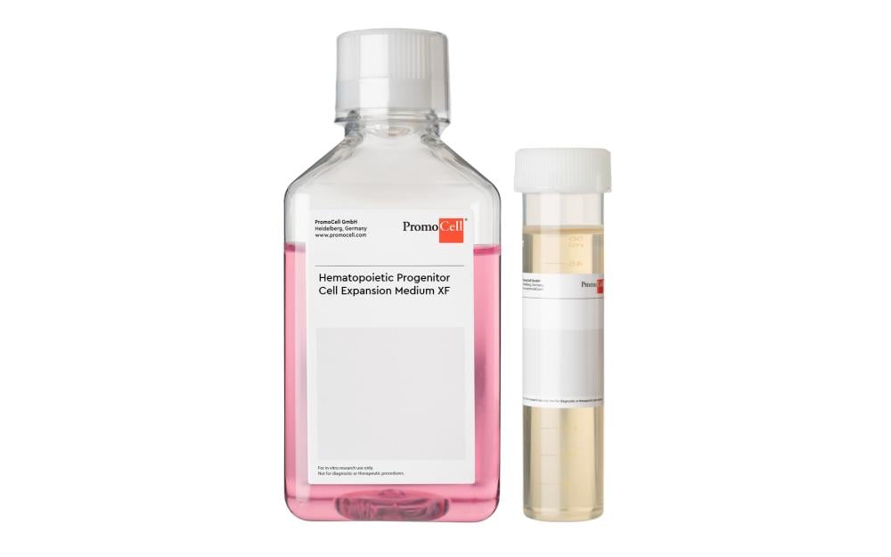
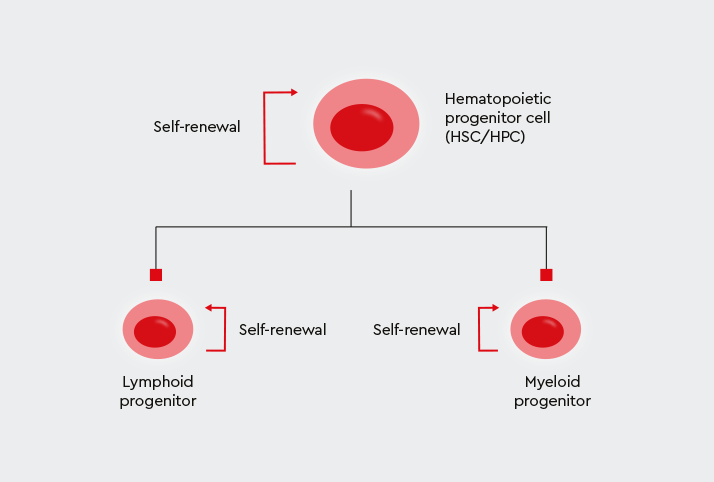
Cytokine Mix E for HPC-Expansion Medium XF
The medium supplemented with Cytokine Mix E is stable for 2 weeks if stored protected from light at 2-8°C.
PromoCell_Best-Practices-in-Cell-Culture
tools-for-respiratory-research_brochure
Standardized culture of assay-ready and fully functional human primary macrophages
Trypsin/EDTA (ready-to-use), 30 ml
DetachKit, 3 x 30 ml
Accutase-Solution
Short description:
Unpack the box and place the T25 flask(s) in the incubator for 3 hrs (closed cap). Then check confluency under the microscope.
When the density is < 70%, aspirate the medium using sterile conditions and add 5-10 ml of the appropriate Growth Medium.
The cells should be subcultured according to the subcultivation protocol given in the cells' Instruction Manual once they have reached > 70 % confluency.
PromoCell_Best-Practices-in-Cell-Culture
tools-for-respiratory-research_brochure
Standardized culture of assay-ready and fully functional human primary macrophages
Human Chondrocytes (HCH)
It usually takes 5 to 8 days to grow our Normal Human Chondrocytes (C-12710) to subconfluency. The number of doublings (PDs) they undergo can be calculated from the number of seeded cells and the cell yield at subconfluency. Generally, when HCH are plated with 10,000 cells/cm² they perform between 1.5 and 2 doublings per passage.